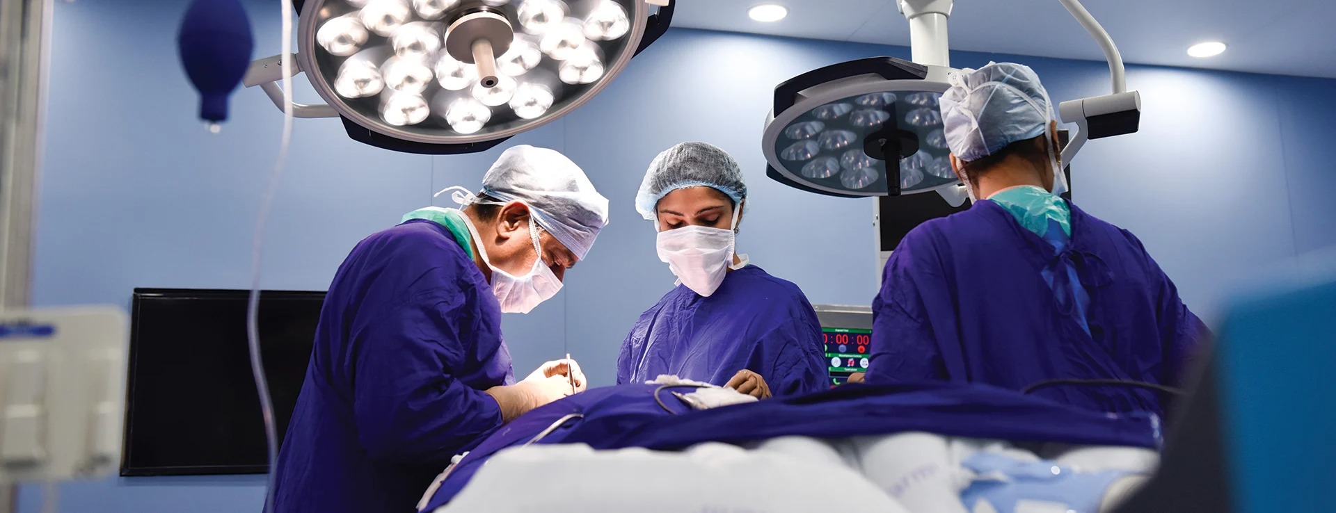
As a truly world-class institution, Kokilaben Hospital offers doctors and patients cutting-edge diagnostic and surgical solutions as well as the latest in IT systems. In many cases, they represent the first of their kind in the region. Some of these wonders of medical and hospital technology include:
Kokilaben Dhirubhai Ambani Hospital introduces the Toumai® Robotic Surgery System. This is the first installation of this groundbreaking surgical system in India. This advanced system brings along a new standard in robotic-assisted minimally invasive surgery, delivering high-end and superior surgical outcomes. Recognized globally with the Red Dot Award and Super AI Leader Award, the Toumai® Endoscopic Multi-Port Surgical System features 3D HD visualization, tremor-filtered wrist instruments, and adaptable multi-arm technology, making this surgical system precise, safe, and efficient in performing complex life-saving surgeries.
Key Benefits for Patients
Kokilaben Dhirubhai Ambani Hospital is proud to unveil the groundbreaking, cutting-edge radiation therapy – the Radixact® Treatment Delivery System, a new age radiation technology system that aims to provide safer, quicker, and precise cancer treatment. This next-generation system by Accuray, Radixact® offers a transformative approach with its helical radiation delivery, which rotates 360 degrees around the patient to precisely target tumors. Equipped with a built-in CT scanner for real-time imaging and the Synchrony® motion tracking technology, this system ensures that the radiation is delivered with perfect precision - only onto the tumour without disturbing any healthy tissues around it. This has indeed set a new benchmark in radiation therapy procedures.
Key Advantages for Patients
Kokilaben Hospital had its first robot few years ago for performing minimally invasive surgery. In a short span of 2 years, our specialists performed over 1000 Robotic Surgeries with successful outcomes. Owing to the overwhelming demand, we launched our second cutting-edge robot da Vinci Xi®.
The hospital now has two da Vinci Surgical systems.
The version of robot has more capabilities than the previous da Vinci Si model. It is optimized for complex, multi-quadrant as well as single-quadrant surgical procedures. Remote access is not the only potential of da Vinci System. The surgeon can also use tiny tools meant for smaller incisions. Visual enhancement is another value-add that provides information to the surgeon that his eyes might not see. It is thus said to be an extension of the surgeon’s eyes and hands.
Here are a few improved facilities provided by the new da Vinci Xi:
The Core Technology that makes da Vinci Xi a better model is:
Kokilaben Hospital has recently launched Western India's first O-arm® O2 Intra-operative 2D/3D Imaging System and Spine Surgery Suite – one of its kinds. Spine Surgery Suite is a unique concept which has some essential and state-of- the-art technologies coming together for getting the best possible outcomes. Technologies that make the Spine Surgery Suite so special are O-arm® O2 Surgical Imaging System, StealthStation® S8 ® Surgical Navigation System, Trios ® Surgical Table and OPMI Pentero 900 Surgical Microscopes.
A Robotic-Driven Upper Limb Orthosis, a Computer Enhanced Rehabilitation Device to improve hand and arm impairment for patients with brain injuries, neurological disorders or strokes
A Robotic-Driven Gait Orthosis, a Computer Enhanced Rehabilitation Device to improve walking in neurologically impaired patients
World over, navigation systems are redefining the way surgeries are performed. They enable surgeons to achieve greater precision by providing electronic guidance using computers, infrared cameras and wireless instruments. Surgeons can effectively make data-driven decisions in the operating room by incorporating the three technological innovations that provide a data-rich, visual surgical environment that helps reduce procedure invasiveness, risk and operative times.
The Novalis TX at Kokilaben Hospital is among the world's first whole radiosurgery systems for indications in the brain, liver, pancreas, prostate and lung. It enables small, deep-seated tumours to be treated by radiation without open surgery, and offers a versatile combination of advanced technologies for the treatment of tumours and other anatomical targets. With this platform, the Kokilaben Hospital offers state-of-the-art, non-invasive treatment for a wide range of cancers and other potentially debilitating conditions, without harming nearby healthy tissue and without involving traditional surgery.
Patients today demand the highest quality of care, including a comfortable exam experience and the assurance that the diagnosis is the most accurate possible. When you combine 3T with a 70 cm open bore, you give them just that. More space limits claustrophobic rejections, fewer patients need to be sedated, and sharper images are captured thanks to less anxiety-related movement. The 3T system at Kokilaben Hospital accommodates patients with special needs and conditions including spinal deformities, respiratory problems and those with pain and mobility issues. It also allows us to expand care to a wider range of patients including obese, paediatric, elderly, ICU patients or those dependent upon medical equipment. The 3T broadens clinical possibilities with easy access in interventional MRI and opportunities to perform more functional studies (using the functional MRI tool).
Kokilaben Hospital is pleased to announce the launch of Mumbai's first Digital PET - CT scan under our Centre for Excellence in Oncology. We are committed to make the fight against cancer easier with the most advanced form of diagnostic facilities. The Beyond Digital PET/CT Biograph Vision 600 PET/CT system, featuring AIDAN technology in India is a revolutionary imaging system. This advanced imaging system sets a new standard in precision oncology with the industry’s sharpest time-of-flight (TOF) resolution at 214 picoseconds. This high level of accuracy enhances performance and reproducibility, which is crucial for tracking disease progression. This high-resolution PET image significantly improves lesion detection and can positively impact health outcomes in cancer patients. With a whole new world of precision, Biograph Vision 600 can make a significant difference in cancer care. Making a profound impact in cancer care and catering to personalized medicine, this system focuses on clinical insights and promotes sustainable health outcomes.
The latest revolution in CT is the new Dual Source CT technology that pioneers new clinical opportunities. Our DSCT is faster than every beating heart, can obtain full cardiac detail at half the dose of radiation, serves as a one-stop diagnosis in emergencies, and goes beyond visualisation with dual energy. Twice as fast as other cardiac CT scanners and less influenced by irregular heart rhythms, it eliminates the need for beta-blockers to slow down the heart rate prior to cardiac scanning. The DSCT at Kokilaben Hospital opens the door to a new world of characterization; visualising the chemical composition of the human body. The result: two spiral data sets acquired in a single scan providing diverse information, which allows us to differentiate, characterise, isolate, and distinguish the imaged tissue and material composition.
Single Photon Emission Computed Tomography (SPECT) is a nuclear medicine imaging technique using gamma rays. SPECT can be used to complement any imaging study where a true 3D representation is needed, such as tumour, infection, thyroid or bone imaging. As SPECT permits accurate localisation in 3D space, it is used to provide information about localised functioning of internal organs; for instance, functional cardiac or brain imaging.
Asia's first Radiosurgery and Radiation Therapy equipment for Cancer Treatment
A new EDGE™ Radiosurgery system from Varian Medical Systems is being installed at Kokilaben Dhirubhai Ambani Hospital for the first time in Asia. The newly-installed device offers a precise, non-invasive alternative to conventional surgery, with no incisions and few of the healing, pain, and recovery issues typically associated with conventional surgery. The installation will make significant difference in precise and quality outcomes in cancer treatment.
ECLIPSE: Planning system for all forms of External beam radiotherapy like 3 D CRT / IMRT
BRACHYVISION: 3D planning system for image-guided brachytherapy which uses 3D data sets such as CT, MR and PET.
We have the latest Olympus EBUS equipment and have been routinely doing EBUS TBNA. EBUS TBNA is minimally invasive and relatively safe procedure for sampling mediastinal and hilar lymph glands and can be offered on day care basis and patients can go home on the same day. It is useful in staging of lung cancer and diagnosis of various non-cancer conditions like TB and Sarcoidosis. Apart from this, we also offer radial probe EBUS for sampling small lesions in the lungs which are not accessible by conventional bronchoscopy or CT guided lung biopsy.
It is a device, which uses ultrasound technology to tell us about the stage of liver disease without biopsy. It also guides about the amount of fat in liver. It is a painless and non-invasive procedure used for diagnosis and treatment of liver disease. Time required for the procedure is short and gives results immediately. It is done on OPD-basis and does not require any fasting on part of patient.
Right from genetic testing to genetic counselling, all under one roof, we at the Molecular Biology and Cytogenetics department of Kokilaben Hospital perform and study genetic tests for the detection of various neurological, cardiovascular and haematological disorders and cancer.
Next-generation sequencing (NGS) has allowed for lower cost, higher-throughput targeted genome sequencing. The MiSeq desktop sequencer provides tailored personalized targeted therapy in cancer patients. It gives genetic insights for diagnosis of complex neurological disease and even modern lifestyle diseases so that people at risk can be identified and managed.
The Applied Biosystems 3500xL Dx Genetic Analyzer is used in establishing genetic diagnosis for patients (eg. Beta thalassemia, Breast cancer- BRCA 1 & 2 gene sequencing, Cystic Fibrosis, neurological disorders-SCAs, HD, etc.) This will help in the management of diseases and offer pre-natal diagnosis in certain medical conditions.
The Applied Biosystems 7500 Fast Dx Real-Time PCR Instrument is a sensitive instrument used to monitor minimal residual disease in patients suffering from CML, AML, ALL and MPD to aid in disease detection and management.
The Blood Bank is equipped with state-of-the-art equipment, which enables use of advanced techniques for processing/screening of blood and its components.
Immunohaematology tests (Blood grouping, Antiglobulin test etc.) are done with latest Column agglutination and Capture technology. Solid-phase red-cell adherence (SPRCA) techniques for platelet serology will be commissioned soon.
Nucleic Amplification Test- We also offer extra layer of blood safety by doing molecular test (NAT) for HIV, Hepatitis B and C which is offered by very few centres in country.Our Urodynamics Laboratory system offers diagnostic services for patients, inpatient, outpatient and a day care basis as required.
The laboratory provides the following services:
The Urodynamics Study Laboratory caters to patients from all age groups ranging from paediatrics to geriatrics. These cater to immuno-compromised and physically challenged individuals as well.
ESWL (Lithotripter) at Kokilaben Dhirubhai Ambani Hospital is highly effective extracorporeal shock wave lithotripsy combined with state-of-the-art endourology – the perfect solution for the entire spectrum of minimally invasive stone therapies.
Kokilaben Dhirubhai Ambani Hospital is now home to highly advanced state-of-the-art biplane catheter laboratory, the first of its kind in the country. In these labs, without performing surgery, physicians (Interventional Neuroradiologists) can remove blood clots (which cause strokes) from the brain and can repair weak arteries (aneurysms) in the brain. At the heart of the laboratory is a unique biplane camera. Aided by a contrast dye injected into the patient's circulatory system, it snaps highly detailed photos of the brain and its maze of blood vessels simultaneously from two distinct angles. Colour pictures of the blood vessels appear in real time on massive monitors where the doctor can rotate the digital data to see the pictures from every conceivable angle prior to skilful intervention. While watching the screen, interventional radiologists insert a catheter via the blood vessels of the groin of the patient, all the way through the abdomen, chest and neck into the brain. Thus, complex abnormalities of the blood vessels of the brain can be treated without surgically opening the skull.
Using microscopic instruments inserted via the catheter, the doctor can remove blood clots, block aneurysms, insert stents or block vessels supplying dangerous masses like cancers and vascular malformations, thereby preventing haemorrhages into the brain and also re-establishing critical normal circulation.
The Artis Q biplane system provides fast, easy handling and streamlined workflow by combining compact and rotating detectors in both planes, including lateral detector rotation that tracks the table as it is tilted. The compact and rotating detectors enable virtually unrestricted patient access while offering outstanding angulation and exceptional coverage capabilities.
Artis Q includes the CARE and CLEAR packages to complement the imaging chain for optimised post-processing and dose reduction. The CARE package helps reduce radiation for the operator and patient. The CLEAR package offers a comprehensive range of applications to enhance image quality. CARE and CLEAR are standard with all Artis Q systems.
Artis Q has Gigalix X-ray tube. This tube has been designed around a unique flat emitter technology that generates powerful short pulses. Compared to filament technology, the higher maximum current of the flat emitter enables CLEAR pulse and enhances image quality in challenging situations such as with obese patients or in steep angulations.
Together with small focal spots, this generates equal image quality with up to 60 per cent less dose
The GIGALIX X-ray tube in the Artis Q product line scores a double win: enhanced image quality at a significantly lower dose for both patients and staff.
In addition to X-ray generation, X-ray detection is crucial for high image quality. The new large detector comprises a 16-bit read-out generating more than 65,000 gray scale values leading to enhanced soft-tissue contrast in 3D imaging, especially at image borders (e.g. close to bones like the skull). Increased scintillator thickness enables higher detective quantum efficiency. This provides imaging excellence even in challenging situations and helps to reduce radiation. The water-cooled design meets high hygienic requirements, especially in hybrid operating rooms.
Introducing “VELYS Robotic System” for the first time in Mumbai, we bring not just navigation to Knee Replacement surgeries but a complete robotic surgery solution. VELYS Robotic-Assisted Solution is a novel, state-of-the-art knee system that simplifies total knee replacement surgery.Robotic-assisted devices can aid your orthopaedic surgeon in performing a precise knee replacement procedure tailored to your unique anatomy. The VELYS Robotic System uses your specific intraoperative data to help surgeons optimise the results of the surgery.
Made with a compact and efficient design, this system integrates into any operating room and is substantially smaller than other common robotic-assisted systems. This leading technology has reported improved patient outcomes, better clinical outcomes, as well as contribute to a shorter hospital stay.
Advantages of VELYS Robotic System:
High Intensity Focused Ultrasound (HIFU) is an advanced procedure to fight prostate cancer. It is a focal ablation treatment that uses high frequency sound waves to destroy prostate cancer cells. The machine gives off sound waves to heat up and destroys the prostate cancer cells. Your doctor may recommend HIFU as a treatment if your cancer is confined to the prostate gland. Use of HIFU is not possible if the cancer has spread to other parts of your body which means it is metastatic or advanced prostate cancer. The most commonly done HIFU treatment is focused towards specific areas of the prostate gland; this is known as focal HIFU. Here are a few key advantages of the HIFU:
Kokilaben Dhirubhai Ambani Hospital launched revolutionary technology, the Arthrex Modular Glenoid System with VIP (Virtual Implant Positioning), for shoulder replacement surgery in India. The Arthrex Modular Glenoid System with VIP marks a substantial leap forward in orthopaedics, providing superior precision, accelerated recovery, and enhanced patient results.
Surgeons plan implant placement precisely before surgery using VIP technology. It creates a virtual shoulder model, ensuring unmatched precision customized to each patient. This approach reduces complications and promotes faster recovery with minimal invasiveness. Patients regain shoulder function swiftly, boosting satisfaction and daily activity resumption.
Kokilaben Dhirubhai Ambani Hospital, one of India's leading super-specialty hospitals and the most accredited hospital in Western India, is equipped with Total Lab Automation (TLA) technologically powered by Roche Diagnostics. This technology is known to improve patient care, with a special emphasis on preventive healthcare.
Total Lab Automation revolutionizes traditional laboratory processes by integrating data analytics, workflow management, and robotics. The platform makes use of cutting-edge robotics, high-definition cameras, customized algorithms, and machine learning to streamline operations, get rid of human error, and produce faster and more accurate results. This multidisciplinary open automation approach has brought advanced technology to patient care.
The Total Lab Automation/Smart Core Lab integrates five major laboratory disciplines - Clinical Chemistry, Toxicology, Immunology, Infectious Serology, and Haematology & Coagulation into one automated track. This helps immensely reduce the turnaround time and provides hassle-free laboratory reports. This system uses centralized real-time monitoring, advanced algorithms, and proactive critical alerts. These state-of-the-art systems provide hospitals with accurate and timely results, thus enhancing overall patient care. The technology's design flawlessly integrates every aspect of laboratory workflows into a user-friendly interface, enhancing both staff safety and satisfaction.
At Kokilaben Dhirubhai Ambani Hospital we take pride in being one of the first centres in the country to provide comprehensive stroke care from acute phase upto neuro rehabilitation. We offer the fastest response time to manage stroke emergencies and provide a door to needle time of less than 60 minutes. Our Centre for Neurosciences provides exceptional care in neurology, neurosurgery and interventional neuroradiology with one of the highest number of endovascular interventions in acute stroke management. We are highly committed to offer world-class stroke care to our patients and continuously upgrade our technologies for the same.
To increase our efficacy in treating stroke patients we are now equipped with a lifesaving advanced software - Rapid AI (Artificial Intelligence) that helps treat stroke emergencies at the earliest. Rapid AI empowers doctors to rapidly assess the severity of the stroke and make faster, more accurate diagnostic and treatment decisions for stroke.
The Centre for Cardiac Sciences at Kokilaben Dhirubhai Ambani Hospital, Mumbai announces the launch of India's first AI-powered, intelligent Artist Icono Cardiac Suite. This pioneering facility integrates the Excimer laser system and advanced imaging technologies to enhance cardiac care precision and patient outcomes. Key features include the "CLEARSTENT live" technology for unparalleled precision in cardiac stenting, and the "syngo DynaCT cardiac" for real-time 3D imaging during valve implantation procedures. The suite is also equipped with the OPTIQ imaging chain, which offers up to 70% dose reduction while optimizing image contrast, and the "structure scout" for high-resolution, material-specific imaging that enhances overall image quality. "Case flows" technology further streamlines workflow, optimizing procedure time for unmatched efficiency in cardiac treatment.


