When the shoulder dislocates, the ball comes off the plate or socket. In many cases the person cannot get the ball back in by themselves, and medical attention is required. The shoulder typically dislocates when the elbow is away from the body and the arm is rotated in a way that the humeral head rolls over the front edge of the socket. Less commonly, the humeral head can be driven off the back of the socket with the arm in front of the body. In either case, the capsule and/or labrum typically tear away from the socket side of the joint when the ball dislocates. In some cases, the capsule and labrum may pull off a piece of bone from the edge of the socket as the ball dislocates.
In some cases the shoulder does not dislocate completely, and the ball spontaneously returns to its normal location after being perched on the rim of the socket. This is called shoulder subluxation and these cases, capsule and/or labral tears may still occur. Other tissues around the shoulder may be injured at the time of dislocation, including the rotator cuff tendons, the biceps tendon, the deltoid muscle and its accompanying nerve, and the cartilage surfaces of the ball and socket joint.
If the dislocation is recognised immediately, it is possible for the person to pull the shoulder back in using their own muscles. After several seconds, however, the pain and muscle spasm from the dislocation typically prevents a person from getting their own ball back in their socket. In the majority of cases, the person seeks medical attention, X-rays are taken to identify the nature of the dislocation, and the shoulder is reduced (put back in) by a medical professional. Pain medicine and sedation may be necessary to get the shoulder back into place. In some cases where the shoulder cannot be put back in, the patient may be taken to the operating room for surgical relocation of the joint. Immediately following successful reduction of the joint, X-rays are obtained to confirm the reduction and the patient is placed in an immobilising device to prevent re-dislocation.
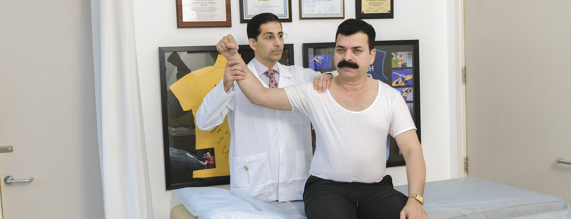
Patients suffering shoulder dislocation are typically placed in an immobilising device for a short period of days to weeks, and early range of motion exercises are initiated to prevent stiffness. In many cases a physical therapist will be consulted to assist with return of motion. Pain medicine and/or anti-inflammatory medicines can be used to decrease pain and swelling.
Later, gradual strengthening exercises are added to return the shoulder to more normal function. Restoring strength to the shoulder also helps prevent re-dislocation. Avoidance of contact sports and other activities where the arm may undergo significant rotation is necessary in the early post-dislocation period to prevent reinjury.
Typically it takes several weeks to return to the routine activities of everyday life and several months to return to heavy lifting and contact sports. In some cases, a shoulder may dislocate more than once, or multiple times, despite adequate management. In these cases, surgery is the best choice.
The younger a person is when they dislocate for the first time the more likely they are to re-dislocate over the course of their life. For example, a 20 year-old who dislocates for the first time has an 80 percent chance of re-dislocation later on. If the shoulder continues to be unstable despite adequate management, it is reasonable to pursue surgical solutions.
In these cases, the capsule and/or labrum are repaired back to the bone from which they tore using sutures and anchors into the bone. In most cases, these repairs are carried out arthroscopically (minimally invasive), but in rare cases ‘open repair’ through a small incision remains the best option. In those cases in which instability occurs repeatedly, surgical solutions have been shown to be nearly 98 percent successful in returning patients to full activity with no limitations.
By increasing the strength of the rotator cuff muscles and avoiding activities which place the shoulder at risk, the likelihood of re-dislocation diminishes. The rotator cuff muscles help squeeze the humeral head into the glenoid socket, thereby increasing the stability. Rehabilitation commonly focuses on the strengthening of these muscles using resistance weights, rubber bands and cables.
If surgical management is undertaken, post-operative rehabilitation begins in the first few days or weeks after surgery. The goal of this therapy is to restore the range of motion and subsequent muscle strength to the shoulder without jeopardising the stability of the recently repaired tissue.
Generally, early exercises are limited to range of motion only, followed several weeks later by strengthening of the rotator cuff after the repaired tissue has had a chance to complete the early healing process. Generally, three months are required for the shoulder to return to normal range of motion and strength, and a subsequent return to full activity.


