Electrical impulses are spontaneously and repetitively generated by the heart and they help optimise the pumping function of the heart. The pattern and timing of these impulses determine the heart rhythm. Derangements in normal rhythm, also referred to as Cardiac Arrhythmias, often impair the heart’s ability to pump enough blood to meet the body’s demands.
Cardiac Electrophysiology is a branch of Cardiology that deals with electrical impulse formation and conduction inside the heart, in other words, the electrical conduction system (wiring) of the heart. The Cardiac Electrophysiologist is a doctor who deals with the disease states resulting from cardiac rhythm abnormalities.
Cardiac Rhythm Abnormalities may be persistent or may be intermittent in nature. If the rhythm abnormality is persistent it can often be diagnosed by obtaining an Electrocardiogram (ECG), which is a way of recording electrical activity of heart by attaching a series of electrodes to the chest wall.
If rhythm abnormality is intermittent it may be more difficult to diagnose and one would need to undertake further investigations, such as 24 hour ambulatory ECG monitoring or even extended periods of heart rate monitoring.After initial consultation and basic investigations, such as ECG and an echocardiogram (an ultrasound scan of the heart – helps in assessing cardiac valves and the pumping function of the heart), the electrophysiologist might commence the patient on medical treatment (called anti arrhythmic medication) or request for an Electrophysiology Study (EPS) for further evaluation of cardiac arrhythmia.
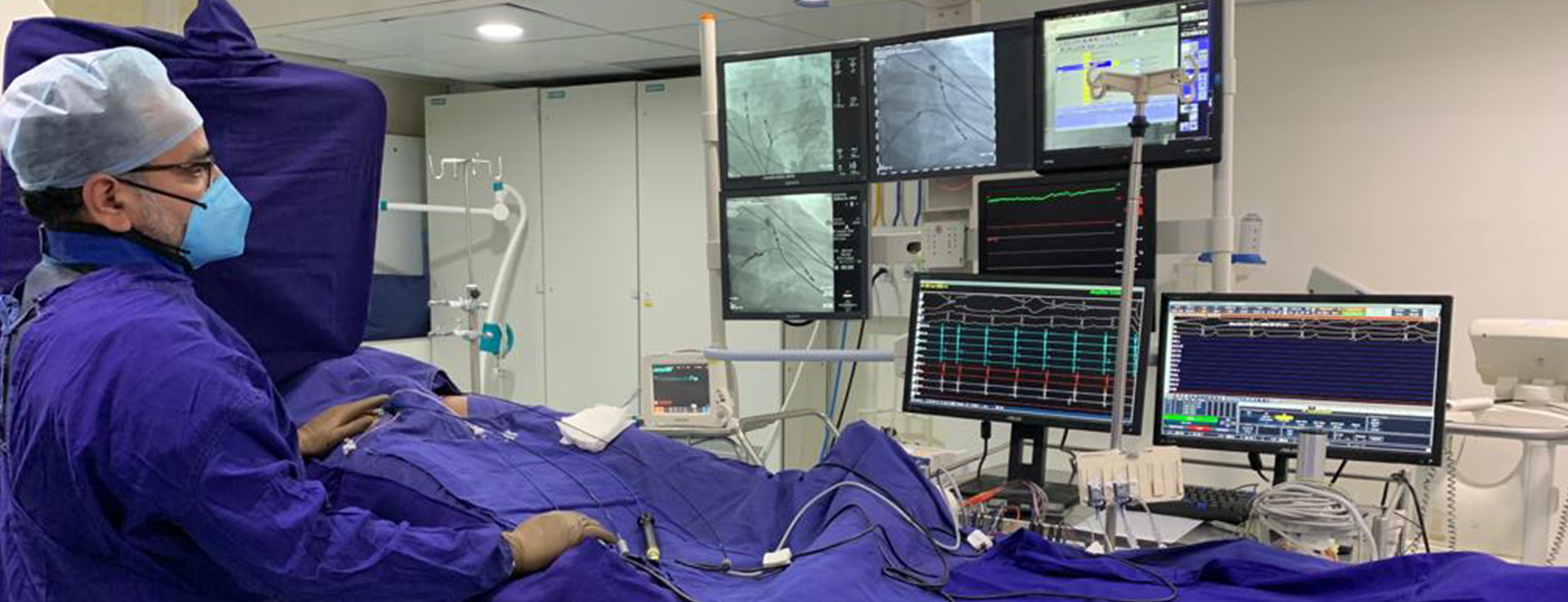
An EPS is the procedure used to evaluate the cardiac electrical system. An electrophysiology study is performed by the Cardiac Electrophysiologist under monitored conditions in a cardiac catheterisation laboratory. A series of thin flexible wires called catheters are introduced into the blood vessels (usually via the groin) and are guided into the heart using fluoroscopy (X-rays). These catheters are positioned in various locations inside the heart. Each catheter has one or more electrodes at its tip and cardiac electrical signals are acquired via these catheters and are displayed on a real time monitor. Electrical signals can be delivered as well as received through the electrode catheters.
During EPS abnormal heart rhythms experienced by the patient are induced by stimulating the heart via the electrodes or by giving cardiac medications via the blood vessels. Once the cardiac arrhythmia is induced, the cause of the abnormality is diagnosed using the signals obtained from various cardiac locations. Often a series of manoeuvres are performed using the electrical signals delivered via the electrodes at the catheter tips.Cardiac arrhythmias could originate from the top chambers of the heart (atrium) in which case they are referred to as supraventricular in origin or they could be from the bottom chambers of the heart which are referred to as being ventricular in origin.
The abnormality associated with the arrhythmia could be a focal point of increased irritability and excitability in heart or alternatively could be due to a re-entrant circuit resulting from abnormal connections (pathways or circuits) in the heart.Once the abnormality is detected your electrophysiologist may treat it using Radio Frequency Ablation (RFA) therapy.
Once a cardiac abnormality is detected (area of concern identified) a special type of catheter called ablation catheter is guided into the heart (via the blood vessels in the groin). The tip of the catheter is positioned at the abnormal point in the cardiac tissue using fluoroscopy and the other end of the catheter is connected to the ablation equipment. Radiofrequency waves are delivered via the tip of the catheter that heats the abnormal cardiac tissue, eventually leading to the destruction of the abnormal focus (or the abnormal pathway). Great care is taken to avoid any damage to the normal healthy cardiac tissue.
EP study is a low risk procedure. Cardiac complications, such as myocardial infarction/stroke or death are extremely rare with incidence of these complications being less than one in thousand procedures.
Risks associated with ablation therapy partly depend on the site of cardiac tissue being ablated. Although infrequent some of the complications associated with ablation therapy include damage to the normal conduction system of the heart necessitating pacemaker implantation and cardiac perforation resulting in seepage of blood into the outer layers of the heart (condition referred to as Cardiac Tamponade).
A normal cardiac impulse originates from sinus node situated in the right top chamber of the heart (right atrium) and spreads across to the left top chamber as well as to the bottom two chambers (ventricles) of the heart. The top and bottom chambers are electrically isolated except for a central region called atri-ventricular node (junction box). Any additional abnormal electrical communications between the top and bottom chambers may result in fast heart rates due to the impulse forming a continuous loop between normal and abnormal pathways and this arrhythmia is referred to as Supraventricular Tachycardia.
The cardiac impulse can at times take an abnormal course in the right atrium resulting in a continuous self perpetuating fast heart rhythm which is referred to as Atrial Flutter.Fast heart rates can also result from rapid firing of cardiac impulses from diseased cardiac tissue in the top chambers often referred to as Atrial Tachycardia.
Abnormal firing of impulses from cells around the draining veins in the left top chamber (left atrium) results in irregular heart rhythm called Atrial Fibrillation.When the fast heart rates are caused due to abnormal impulse generation in the lower chambers of the heart (ventricles) it is referred as Ventricular Tachycardia.
All of these above mentioned rhythm abnormalities can be diagnosed and successfully treated by performing EPS and ablation therapy.Patients who have experienced ventricular tachycardia or who are at high risk of having ventricular tachycardia are advised implantation of an Automatic Internal Cardiac Defibrillator. A cardiac defibrillator is very similar to a pacemaker but has additional capabilities. The cardiac defibrillator device detects a fast heart rhythm and automatically delivers a small electrical shock to the heart to restore normal rhythm.

Electrophysiology (EP) studies and ablation therapy for atrial and ventricular arrhythmia’s including atrial fibrillation and ventricular tachycardia. Implantation of cardiac devices including pacemakers for treatment of slow heart rhythms and advanced cardiac device (defibrillators, cardiac resynchronization therapy devices (CRT) and His bundle pacing) for patients with advanced heart failure.


