
Obsessive-compulsive disorder is one of the most frequently misrepresented mental health conditions in everyday language. It is routinely described as a preference for tidiness, a tendency toward perfectionism, or a harmless personality quirk — none of which bear any clinical resemblance to what OCD actually is.
OCD is a serious, chronic mental health condition in which a person becomes trapped in a cycle of unwanted, distressing thoughts and repetitive behaviours they feel compelled to perform — even when those thoughts make no logical sense. The distress it causes is real, and for many people, it consumes hours of every day. Left unaddressed, it progressively affects relationships, work, and quality of life.
This article explains what OCD actually is, how it works, what drives it, and what effective treatment looks like.
What Is OCD?
OCD, in clinical terms, refers to a chronic mental health condition characterised by two defining features: obsessions and compulsions. These two components interact in a self-reinforcing cycle that, without appropriate treatment, tends to worsen over time.
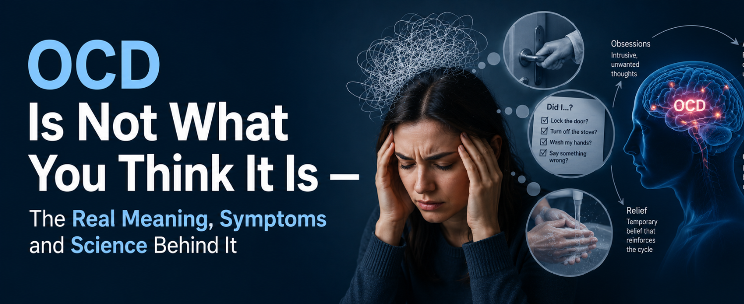
Obsessions are unwanted, intrusive thoughts, images, or urges that enter the mind repeatedly and cause significant anxiety or distress. They are not chosen, not welcome, and not reflective of the person’s actual desires or values. A person with OCD does not want these thoughts — they experience them as invasive and deeply disturbing.
Compulsions are repetitive behaviours or mental acts performed in response to obsessions, with the aim of reducing the anxiety they generate or preventing a feared outcome. The relief compulsions provide is real but temporary. Within a short time, the obsession returns — and the compulsion must be repeated. This is the core of OCD.
What is OCD if not simply anxiety? It is distinguished from general anxiety by the presence of this specific obsession-compulsion cycle, by the ego-dystonic nature of the obsessions, they feel foreign and contrary to the person’s values, and by the time the cycle consumes. DSM-5 criteria require that obsessions and compulsions take up more than one hour per day and cause clinically significant distress or functional impairment.
OCD affects people across all ages, cultures, and levels of education. It commonly begins in childhood, adolescence, or early adulthood. Without treatment, it typically follows a chronic course with periods of exacerbation and partial remission.
The Obsession-Compulsion Cycle — How OCD Actually Works
Understanding the obsession-compulsion cycle is fundamental to understanding why OCD is so difficult to manage without professional support.
The cycle typically operates as follows:
- Trigger — an internal thought, external situation, or sensory input activates the obsession. The trigger itself may be entirely ordinary — a door handle, a sharp object, or a passing thought.
- Obsession — the intrusive thought, image, or urge takes hold and generates intense anxiety or discomfort.
- Interpretation — the person interprets the obsession as meaningful, threatening, or morally significant, amplifying the distress.
- Compulsion — a behaviour or mental ritual is performed to reduce anxiety, neutralise the thought, or prevent the feared outcome.
- Temporary relief — the anxiety decreases briefly, reinforcing the compulsion as a coping mechanism.
- Return of obsession — the obsession returns, often with greater intensity, and the cycle begins again.
Each compulsion prevents the brain from learning that the feared outcome would not have occurred and from experiencing the natural reduction in anxiety that comes from tolerating uncertainty. Over time, obsessions become more frequent, compulsions more elaborate, and the condition more entrenched.
OCD Symptoms — What Obsessions and Compulsions Actually Look Like
OCD symptoms are far more varied than the stereotype of handwashing or tidiness suggests. The content of obsessions reflects whatever a person holds most important or most fears — which is part of what makes them so distressing.
Common Obsession Themes
- Contamination — fear of germs, disease, bodily fluids, or environmental contaminants, even when exposure is objectively minimal
- Harm — intrusive thoughts about accidentally or intentionally hurting oneself or others; fear of being responsible for a catastrophic event
- Symmetry and exactness — a need for things to be arranged or completed in a specific way until they feel “just right”; intense discomfort when they are not
- Unwanted sexual or violent thoughts — disturbing intrusive thoughts completely contrary to the person’s values; distressing precisely because of their nature
- Religious and moral scrupulosity — excessive concern about sin, blasphemy, or having committed a moral wrong; relentless doubt about one’s own character
- Relationship OCD — obsessive doubt about whether one truly loves a partner or whether the relationship is right
Common Compulsion Patterns
- Washing and cleaning — handwashing or cleaning objects and surfaces beyond what hygiene requires
- Checking — repeatedly verifying locks, appliances, or light switches; seeking repeated reassurance that harm has not occurred
- Arranging and ordering — placing objects in specific configurations; repeating actions until they feel symmetrically correct
- Mental rituals — counting, praying, repeating specific words internally, or mentally reviewing events to neutralise a thought
- Avoidance — restructuring daily life to avoid triggers; functionally a compulsion because it maintains the cycle while providing short-term anxiety relief
The Types of OCD Most People Have Never Heard Of
OCD is not a single uniform presentation. Several subtypes are clinically recognised, many of which bear no resemblance to the public image of the condition.
Pure O (Purely Obsessional OCD) A form of OCD in which compulsions are primarily mental rather than behavioural — internal reassurance-seeking, mental reviewing, or thought suppression. The absence of visible rituals leads to significant underdiagnosis.
Scrupulosity OCD Centred on religious or moral perfectionism. The person is tormented by doubt about whether they have sinned or acted immorally — regardless of reassurance or evidence to the contrary.
Relationship OCD (ROCD) Obsessive doubt focused on the validity of a romantic relationship. The person is consumed by questions about whether they truly love their partner, despite having no objective reason to doubt it.
Somatic OCD Excessive preoccupation with body sensations or automatic bodily functions such as breathing, swallowing, heartbeat — that becomes conscious and distressing through focused attention.
PANDAS/PANS-related OCD In some children, OCD symptoms develop or worsen abruptly following a streptococcal infection such as strep throat. This form — Paediatric Autoimmune Neuropsychiatric Disorders Associated with Streptococcal infections — is thought to involve an autoimmune mechanism in which antibodies directed at the streptococcus bacterium cross-react with brain tissue. Assessment of this form may require collaboration between psychiatry and the department of rheumatology or immunology, given its autoimmune underpinning.
What Causes OCD — The Science Behind It
What causes OCD is not a single factor. It arises from the interaction of neurobiological, genetic, and environmental influences.
Neurological basis Neuroimaging consistently identifies abnormalities in circuits connecting the orbitofrontal cortex, anterior cingulate cortex, thalamus, and basal ganglia — a loop involved in decision-making, error detection, and behavioural inhibition. In OCD, this circuit appears hyperactive, generating persistent “error signals” that the brain interprets as requiring a corrective response — the compulsion. Serotonin and glutamate neurotransmitter systems are both implicated in this dysregulation.
Genetic factors OCD runs in families. Having a first-degree biological relative with OCD significantly increases an individual’s risk, particularly if the relative’s onset was in childhood or adolescence. Twin studies confirm a meaningful heritable component, though no single gene has been identified as the definitive cause.
Psychological and environmental factors Early life experiences, including trauma, abuse, neglect, or environments characterised by excessive responsibility or perfectionism — can shape the cognitive patterns that contribute to OCD vulnerability. Stressful life events frequently trigger or exacerbate OCD in individuals with a pre-existing neurobiological predisposition.
OCD Diagnosis — How It Is Formally Assessed
OCD diagnosis is made by a psychiatrist or clinical psychologist using criteria from the Diagnostic and Statistical Manual of Mental Disorders, 5th Edition (DSM-5). There is no blood test or imaging study that confirms OCD.
Diagnostic criteria require:
- The presence of obsessions, compulsions, or both
- Those symptoms consume more than one hour per day or cause significant distress or functional impairment
- That symptoms are not attributable to substances, medications, or another medical condition
- That the presentation is not better explained by another psychiatric condition
Clinical assessment typically includes a structured diagnostic interview, standardised rating scales such as the Yale-Brown Obsessive Compulsive Scale (Y-BOCS), and evaluation of symptom severity and functional impact.
A critical clinical note: the average delay between OCD symptom onset and first clinical contact is seven to ten years. Shame, misattribution of symptoms, and a lack of awareness that what a person is experiencing is OCD, rather than personal weakness, are the primary drivers of this delay. Earlier treatment consistently produces better long-term outcomes.
OCD Treatment — What Actually Works
OCD treatment has a well-established evidence base. While OCD is a chronic condition in most cases, meaningful symptom reduction and functional recovery are achievable for the majority of patients.
Exposure and Response Prevention (ERP) ERP is the gold standard psychological treatment for OCD. It is a structured form of cognitive behavioural therapy in which the patient is progressively exposed to anxiety-provoking stimuli related to their obsessions, while being supported in refraining from the compulsive response. Over repeated exposures, the anxiety generated by the obsession naturally decreases without the compulsion — breaking the reinforcement cycle and restoring the brain’s ability to tolerate uncertainty.
Pharmacotherapy Selective serotonin reuptake inhibitors (SSRIs) — including fluoxetine, sertraline, fluvoxamine, and paroxetine — are the first-line pharmacological agents for OCD. They reduce the intensity and frequency of obsessions and compulsions in a significant proportion of patients. OCD typically requires higher doses than those used for depression, and response develops over a longer timeframe. Clomipramine, a tricyclic antidepressant with strong serotonergic activity, is used when SSRIs are insufficient.
Combined treatment The combination of ERP and pharmacotherapy produces better outcomes than either approach alone for most patients with moderate to severe OCD.
Advanced interventions for treatment-resistant OCD For patients who do not respond adequately to multiple treatment trials, augmentation strategies including antipsychotic addition, intensive ERP programs, transcranial magnetic stimulation (TMS), and, in carefully selected cases, deep brain stimulation are considered. The best neuro rehabilitation centre in India, with expertise in treatment-resistant psychiatric conditions and neurostimulation, provides the most structured pathway for patients whose OCD has not responded to standard treatment.
For initial assessment, accurate diagnosis, and development of a comprehensive OCD treatment plan, consultation with the best psychiatrist in India with specialist expertise in OCD and anxiety disorders is the most important first step.
Common OCD Myths — Formally Debunked
Myth: OCD is about being clean or organised OCD can involve contamination themes, but the majority of OCD presentations have nothing to do with cleanliness. Harm, religion, relationships, intrusive thoughts, and somatic preoccupations are equally common — and far less visible.
Myth: People with OCD can just stop if they try hard enough Compulsions are not choices. They are driven by intense anxiety and reinforced by temporary relief. Willpower alone does not break the obsession-compulsion cycle — structured treatment does.
Myth: OCD is caused by poor parenting or weak character OCD is a neurobiological condition with identifiable brain circuit abnormalities and a genetic component. It is not a reflection of character, willpower, or upbringing.
Myth: OCD always involves visible rituals Many people with OCD perform purely mental compulsions — internal reviewing, reassurance-seeking, thought suppression — with no visible behavioural signs. These presentations are frequently missed or misdiagnosed.
Myth: OCD is rare OCD affects approximately one to two percent of the global population, making it one of the more prevalent mental health conditions worldwide. It is underdiagnosed, but not uncommon.
Conclusion
OCD is not a personality trait. It is not a preference for tidiness, a tendency toward perfectionism, or a sign of being detail-oriented. It is a clinically significant, neurologically grounded condition that traps individuals in a cycle of distressing intrusive thoughts and time-consuming compulsive behaviours — a cycle that, without appropriate treatment typically intensifies over time.
The most important clinical message is this: OCD is treatable. Exposure and Response Prevention therapy, pharmacological intervention, and where needed, advanced neurostimulation approaches produce meaningful recovery for the majority of patients. The barrier is rarely the availability of treatment — it is the delay in reaching it.
If you or a family member is experiencing intrusive thoughts, repetitive behaviours, or a pattern of anxiety-driven rituals that is consuming time and interfering with daily life, a clinical evaluation is the right next step. At Kokilaben Dhirubhai Ambani Hospital, our psychiatry team provides specialist assessment and evidence-based management for OCD across all age groups and presentations. Book a consultation today.
Frequently Asked Questions
Q1. Can OCD develop suddenly in adulthood with no prior history — and what usually triggers it?
Yes. While OCD most commonly begins in childhood or early adulthood, new onset in later adulthood does occur. Common triggers include significant life stressors, bereavement, childbirth (postpartum OCD), major health events, and in some cases, neurological illness or infection.
Q2. Is OCD more common in men or women — and does it present differently across genders?
OCD affects men and women at roughly equal rates overall. Men tend to develop it earlier — in childhood or adolescence — while women more commonly develop it in early adulthood. Contamination and checking themes are somewhat more prevalent in women; symmetry and forbidden thought themes are somewhat more common in men.
Q3. Can OCD go into remission on its own — and what causes it to flare up?
Partial remission can occur, particularly in adolescence, but full spontaneous resolution without treatment is uncommon. Flare-ups are typically associated with stress, major life transitions, sleep disruption, illness, or hormonal changes.
Q4. How does OCD affect children — and how do parents recognise it versus normal childhood anxiety?
OCD in children often presents as avoidance of specific situations, elaborate bedtime rituals, repeated reassurance-seeking, or sudden deterioration in school performance. Normal childhood anxiety is typically transient and proportionate. OCD is persistent, time-consuming, and significantly distressing, and the child is usually aware that their thoughts or behaviours are not quite right.
Q5. Is there a connection between OCD and gut health — and does nutrition affect OCD severity?
Emerging research suggests a gut-brain axis connection, with gut microbiome composition potentially influencing serotonin production and neuroinflammatory pathways relevant to OCD. While nutrition is not a treatment for OCD, diets that support gut health and reduce systemic inflammation may complement clinical management. This remains an active area of research rather than established clinical guidance.