Archive for the ‘ Cancer ’ Category

What Is Targeted Therapy for Cancer? A Complete Guide

Friday, June 12th, 2026

Cancer treatment has changed considerably over the past two decades. Where chemotherapy and radiation were once the primary tools available, oncology now has access to a growing class of treatments designed with far greater precision. Targeted therapy for cancer is among the most significant of these advances, not because it replaces other treatments, but because it approaches cancer in a fundamentally different way.

Unlike chemotherapy, which acts broadly on all rapidly dividing cells, targeted therapy identifies and acts on specific molecular structures that drive cancer growth. The result is a treatment approach that is, in many cases, more precise, better tolerated, and capable of producing meaningful outcomes even in cancers that have not responded to conventional treatment. Understanding what targeted therapy is, how it works, and who it is appropriate for is increasingly important for patients and families navigating a cancer diagnosis.

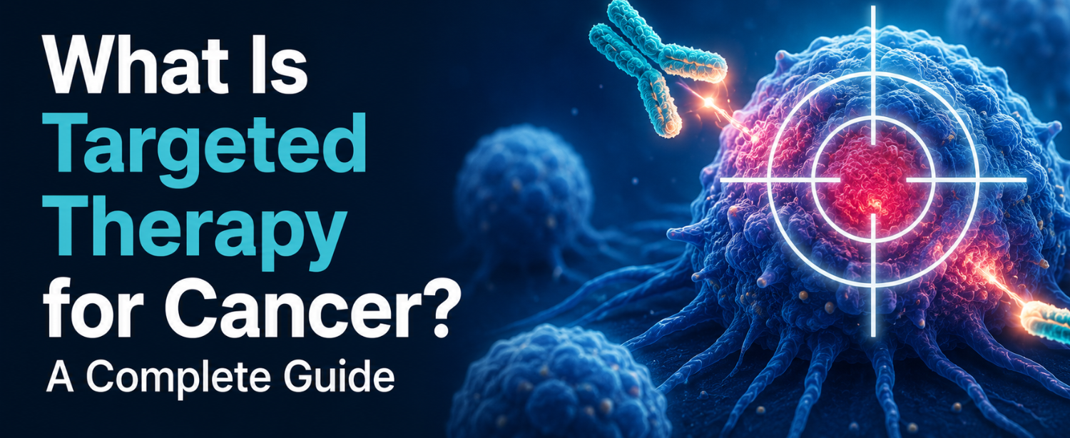
What Is Targeted Therapy in Cancer?

Targeted therapy is a form of cancer treatment that acts on specific proteins, genes, or cellular pathways that cancer cells depend on to grow, divide, and survive. These molecular targets are identified through testing of the tumour’s genetic and molecular profile, a process called biomarker testing or molecular profiling.

Cancer cells develop because of changes, mutations, in the DNA of normal cells. These mutations produce abnormal proteins that drive uncontrolled cell growth. Targeted therapy for cancer is designed to interfere with these specific abnormal proteins, blocking the signals that allow the tumour to grow and spread.

This is what distinguishes targeted therapy from conventional chemotherapy. Chemotherapy is broadly cytotoxic, it disrupts cell division across the board, affecting both cancerous and healthy cells. Targeted therapy acts selectively on the molecular mechanisms that are specifically active in cancer cells, which is why it is often described as a component of precision medicine or personalised cancer treatment.

Not every patient with a given cancer type will be a candidate for the same targeted agent. Eligibility depends on whether the individual tumour carries the molecular target that the drug is designed to act on. Two patients with the same cancer diagnosis may receive entirely different targeted therapies, or one may receive targeted therapy while the other does not, based on what the molecular testing reveals.

How Does Targeted Therapy Work?

At its core, targeted therapy for cancer works by disrupting the specific biological processes that cancer cells rely on. The mechanisms through which this occurs include:

Blocking growth signals Normal cells divide only when they receive signals instructing them to do so. These signals bind to proteins on the cell surface called receptors. In many cancers, the receptors or the signalling pathways they activate are abnormally overactive, causing cells to divide continuously without proper regulation. Targeted drugs can block these receptors or interfere with the downstream signalling proteins, interrupting the growth signal.

Inhibiting blood vessel formation (angiogenesis) Tumours require a blood supply to grow beyond a certain size. They achieve this by releasing signals that stimulate the formation of new blood vessels — a process called angiogenesis. A class of targeted agents known as angiogenesis inhibitors block these signals, depriving the tumour of the vascular supply it needs to grow and sustain itself.

Triggering cancer cell death (apoptosis) In healthy cells, a built-in process of programmed cell death removes damaged or abnormal cells before they can proliferate. Many cancer cells evade this process. Some targeted therapies restore or directly activate the apoptotic pathway, causing cancer cells to self-destruct.

Delivering cytotoxic agents directly to cancer cells A subset of targeted treatments, called antibody-drug conjugates (ADCs), combine a monoclonal antibody with a chemotherapy or toxin payload. The antibody homes in on the target protein on the cancer cell’s surface and delivers the cytotoxic agent directly to the cell, minimising exposure to surrounding healthy tissue.

Supporting immune recognition Certain targeted therapies mark cancer cells with molecules that make them more visible to the immune system, facilitating their destruction by the body’s own immune response. This is distinct from immunotherapy, though the two share some overlapping mechanisms.

Types of Targeted Therapy

Targeted therapy for cancer encompasses several categories of drugs, each with a distinct mechanism and application.

Small-Molecule Inhibitors

These are drugs small enough to enter cells directly and interfere with proteins that function inside the cell. They are commonly used when the molecular target is located within the cell rather than on its surface. Examples include tyrosine kinase inhibitors (TKIs) such as imatinib (used in chronic myelogenous leukaemia), erlotinib and gefitinib (used in certain lung cancers), and lapatinib (used in HER2-positive breast cancer). PARP inhibitors, used in BRCA-mutated breast and ovarian cancers, also fall within this category.

Monoclonal Antibodies

These are laboratory-produced proteins designed to attach to specific targets on the surface of cancer cells. Once attached, they can:

  • Block growth factor receptors from receiving signals
  • Flag the cancer cell for destruction by the immune system
  • Deliver a toxic payload directly to the target cell

Well-known examples include trastuzumab (Herceptin), used in HER2-positive breast and gastric cancer; rituximab, used in B-cell lymphomas; bevacizumab, an angiogenesis inhibitor used in colorectal, lung, and other cancers; and cetuximab, used in certain colorectal and head and neck cancers.

Antibody-Drug Conjugates (ADCs)

ADCs are an advancing category that links a monoclonal antibody to a chemotherapy agent. The antibody delivers the chemotherapy directly to cells carrying the specific target protein. This approach improves the precision of chemotherapy delivery and is an active area of drug development in breast, bladder, and gastric cancer.

Proteasome Inhibitors

These drugs block the proteasome, a cellular structure responsible for breaking down damaged or abnormal proteins. When the proteasome is inhibited, these proteins accumulate and disrupt the cancer cell’s normal function, ultimately causing cell death. Bortezomib and carfilzomib are examples used in multiple myeloma.

mTOR Inhibitors and CDK Inhibitors

mTOR inhibitors target a protein that regulates cell growth and metabolism, used in hormone receptor-positive breast cancer and certain kidney cancers. CDK 4/6 inhibitors, such as palbociclib and ribociclib, block proteins that drive cell cycle progression and are a standard of care in advanced hormone receptor-positive breast cancer.

Which Cancers Can Be Treated with Targeted Therapy?

Targeted therapy for cancer is now approved and used across a wide range of cancer types. The applicability depends on whether the tumour carries the relevant molecular target.

Breast cancer HER2-positive breast cancer is treated with trastuzumab, pertuzumab, and newer ADCs such as trastuzumab deruxtecan. Hormone receptor-positive advanced breast cancer is treated with CDK 4/6 inhibitors and mTOR inhibitors. BRCA-mutated breast cancer is treated with PARP inhibitors.

Lung cancer Non-small cell lung cancer (NSCLC) with EGFR mutations, ALK rearrangements, ROS1 rearrangements, and other driver mutations is treated with targeted tyrosine kinase inhibitors. Molecular testing is now standard before first-line treatment decisions in NSCLC.

Leukaemia and lymphoma Chronic myelogenous leukaemia (CML) with the BCR-ABL fusion gene was one of the first cancers successfully treated with targeted therapy — imatinib transformed outcomes in this disease. Rituximab is a standard component of treatment in B-cell non-Hodgkin lymphoma.

Colorectal cancer Anti-VEGF agents (bevacizumab) and anti-EGFR agents (cetuximab, panitumumab) are used in RAS wild-type metastatic colorectal cancer.

Melanoma BRAF-mutated melanoma is treated with BRAF and MEK inhibitors, which have produced significant improvements in outcomes for advanced disease.

Gastrointestinal stromal tumours (GIST) KIT and PDGFRA mutations in GIST are directly targeted by imatinib and related agents.

Other cancers Targeted agents are also approved or under evaluation in thyroid cancer, kidney cancer, ovarian cancer, bladder cancer, gastric and gastroesophageal junction cancer, and multiple myeloma, among others.

How Is Targeted Therapy Administered?

The mode of administration varies depending on the specific drug and the cancer being treated.

Oral tablets or capsules Many small-molecule targeted agents are taken as daily oral medications. This allows outpatient treatment without the need for infusion visits, though adherence and monitoring of side effects remain important.

Intravenous infusion Monoclonal antibodies are typically administered as intravenous infusions in a clinical setting. Infusion frequency varies by drug — some are given weekly, others every three weeks or monthly — and infusion sessions may take between 30 minutes and several hours.

Subcutaneous injection Some monoclonal antibodies, such as certain formulations of trastuzumab, are available as subcutaneous injections, which are faster to administer and do not require intravenous access.

Targeted therapy may be used as the primary treatment, in combination with chemotherapy or hormonal therapy, before surgery (neoadjuvant), after surgery (adjuvant) to reduce recurrence risk, or in the metastatic setting to control disease spread.

What Are the Side Effects of Targeted Therapy?

Targeted therapy side effects are generally distinct from those of chemotherapy, though they are not absent. Because targeted drugs act on specific molecular pathways, the side effect profile reflects the tissues and systems in which those pathways are also active in normal biology.

Common targeted therapy side effects include:

  • Skin and nail changes — rash, dry skin, hand-foot syndrome (redness, peeling, and tenderness of the palms and soles), and nail changes are among the most frequently reported side effects, particularly with EGFR inhibitors
  • Hypertension — elevated blood pressure is a well-recognised side effect of angiogenesis inhibitors such as bevacizumab, requiring active monitoring and management
  • Fatigue — common across most targeted agents, though generally less severe than chemotherapy-related fatigue in many patients
  • Gastrointestinal effects — diarrhoea, nausea, and mouth sores occur with several classes of targeted agents, including CDK inhibitors and some TKIs
  • Liver toxicity — elevated liver enzymes are monitored through regular blood tests during targeted therapy; significant hepatotoxicity requires dose adjustment or cessation
  • Wound healing impairment — angiogenesis inhibitors can impair surgical wound healing and typically require a washout period before and after surgical procedures
  • Cardiac effects — certain agents, particularly trastuzumab and some TKIs, require baseline and periodic cardiac monitoring due to their potential effect on heart function
  • Haematological changes — reduced white blood cell or platelet counts occur with some targeted agents, particularly CDK 4/6 inhibitors, requiring dose modifications in some patients
  • Infusion reactions — for intravenously administered monoclonal antibodies, infusion-related reactions including fever, chills, and flushing can occur, particularly with the first administration

Managing targeted therapy side effects requires active collaboration between the patient and the oncology team. Many side effects are manageable with appropriate supportive care, dose adjustments, or temporary treatment interruptions. Patients should report new or worsening symptoms promptly rather than tolerating them in silence.

What to Expect During Treatment at Kokilaben Dhirubhai Ambani Hospital

At Kokilaben Dhirubhai Ambani Hospital, the approach to targeted therapy for cancer begins before treatment — with a thorough molecular and pathological evaluation of the tumour to establish eligibility for specific agents and to guide the most appropriate treatment strategy.

Diagnosis and molecular profiling All patients being considered for targeted therapy undergo comprehensive biomarker testing. This includes immunohistochemistry (IHC), fluorescence in situ hybridisation (FISH), and next-generation sequencing (NGS) where indicated, to identify the specific molecular targets present in the tumour. The results of this profiling directly inform the treatment plan.

Multidisciplinary tumour board review Each case is reviewed by a multidisciplinary oncology team comprising medical oncologists, surgical oncologists, radiation oncologists, pathologists, and radiologists. This collaborative review ensures that the decision to use targeted therapy is made in the context of the complete clinical picture.

Treatment planning and administration Oral targeted agents are initiated with a structured patient education session covering dosing, administration, potential side effects, and when to seek urgent review. Intravenous targeted therapies are administered in KDAH’s dedicated oncology day care unit under nursing supervision.

Monitoring and response assessment Regular blood tests, imaging, and clinical assessments track treatment response and detect toxicities early. Treatment adjustments are made based on objective response criteria and tolerability.

For families seeking a comprehensive cancer care pathway — from diagnosis through molecular testing, treatment, and supportive care — the best cancer hospital in India combines the full spectrum of oncological expertise under one roof. Our medical oncology department is equipped with the latest targeted agents and the diagnostic infrastructure required to deploy them precisely. For individuals seeking an initial consultation or a second opinion on a cancer diagnosis, a cancer specialist doctor at Kokilaben Dhirubhai Ambani Hospital can provide a thorough evaluation and a clearly structured management plan.

Conclusion

Targeted therapy for cancer represents a clinically meaningful advance in the treatment of many cancer types. By acting on the specific molecular drivers of individual tumours rather than broadly suppressing cell division, it has improved outcomes, expanded treatment options for cancers previously resistant to chemotherapy, and in many cases reduced the severity of treatment-related side effects.

It is not a universal solution. Eligibility depends on the molecular characteristics of the tumour, and resistance remains a clinical challenge. But for patients whose cancers carry the relevant targets, it has fundamentally changed what is possible in terms of disease control, quality of life during treatment, and long-term outcomes.

At Kokilaben Dhirubhai Ambani Hospital, our oncology programme integrates molecular diagnostics, multidisciplinary expertise, and access to current targeted agents to ensure that every patient receives a treatment plan that reflects the specific biology of their disease. If you or a family member has received a cancer diagnosis and would like to understand whether targeted therapy is an appropriate option, we encourage you to book a consultation with our oncology team today.

Frequently Asked Questions

Is targeted therapy the same as immunotherapy? 

No. Targeted therapy acts on specific proteins that drive cancer cell growth. Immunotherapy works by activating or modifying the immune system to recognise and destroy cancer cells. Some drugs have overlapping mechanisms, but they are distinct treatment categories.

Can targeted therapy cure cancer? 

In a small number of cases, such as certain leukaemias and GIST, targeted therapy can produce deep, sustained remissions equivalent to a functional cure. In most solid tumours, particularly in advanced stages, it controls disease and extends survival rather than curing it outright.

How long does targeted therapy treatment last? 

Duration varies by cancer type, treatment intent, and response. Some patients take oral targeted agents for years. Others receive treatment for a defined number of cycles. Treatment continues as long as the cancer is responding and side effects are tolerable.

What happens if targeted therapy stops working? 

Cancer cells can develop resistance mechanisms over time. When this occurs, oncologists may switch to an alternative targeted agent, a different drug class, or a combination strategy. Repeat molecular testing of the tumour is often performed to identify the resistance mechanism and guide the next treatment decision.

Is targeted therapy available in India? 

Yes. A growing number of targeted agents are now approved by the Central Drugs Standard Control Organisation (CDSCO) and available in India. Leading cancer centres including KDAH have access to molecular testing infrastructure and a range of targeted therapies across breast, lung, colorectal, haematological, and other cancers.

Prostate Cancer: Symptoms, Causes, Diagnosis & Treatment Every Indian Man Should Know

Sunday, April 12th, 2026

Prostate cancer affects about one in eight men worldwide. In India, awareness of prostate health is low, and around 85% of cases are detected at Stage 3 or 4, compared with mostly early detection in Western countries. This late diagnosis makes awareness, screening, and timely treatment especially important. Prostate cancer affects approximately one in eight men, yet awareness of prostate health in India remains limited, and many men only learn about the gland when a problem arises. 

Although incidence is currently lower than in many Western countries, cases are rising, and late diagnosis often leads to more complex treatment and less predictable outcomes. Timely detection, regular screening, and reliable information can make the difference between a manageable condition and a life‑threatening disease.

What Is Prostate Cancer?

Prostate cancer meaning in simple terms, is an uncontrolled growth of abnormal cells in the prostate gland, a small, walnut-sized organ situated just below the urinary bladder in males. The prostate produces seminal fluid that nourishes and transports sperm, and it surrounds the urethra, the tube through which urine flows out of the body.

What is prostate cancer at the cellular level? It begins when the DNA inside prostate cells mutates, causing them to divide uncontrollably instead of following the normal cycle of growth and death. Over time, these rogue cells form a tumour. In many men, this tumour grows slowly and stays confined to the prostate for years. In others, it can be aggressive, spreading to nearby tissues, lymph nodes, bones, and distant organs.

Most cases are a type called adenocarcinoma, which originates in the gland cells. Rarer forms, such as small cell carcinoma or sarcoma, tend to behave more aggressively and are treated differently.

In India, approximately 85% of prostate cancer cases are detected at Stage 3 or 4, a sharp difference to the US, where most cases are found early. This makes awareness, screening, and timely diagnosis critically important for every Indian man. 

Prostate Cancer Symptoms And What to Watch For

One of the most challenging aspects of prostate cancer symptoms is that early-stage disease often causes no symptoms at all. The prostate can harbour a growing tumour for years without causing any noticeable symptoms. This is precisely why routine screening saves lives.

These symptoms can be broadly classified into two categories.

Urinary Symptoms

As the prostate wraps around the urethra, any abnormal growth, cancerous or not, can affect urination. Key urinary symptoms to look out for include:

  • Difficulty starting urination, even when one feels the urge strongly
  • A weak, slow, or interrupted urine stream
  • Frequent urination, especially waking up multiple times at night
  • A feeling that the bladder never fully empties
  • Burning or pain during urination
  • Blood in urine (hematuria) or blood in semen (hematospermia)
  • Painful ejaculation
  • Urine leakage or dribbling after finishing urination

Important to note: these same symptoms can also be caused by Benign Prostatic Hyperplasia (BPH) which is a non-cancerous enlargement of the prostate or prostatitis (prostate inflammation). The presence of these symptoms is not a definitive diagnosis of cancer, but they always warrant evaluation by a urologist or oncologist. 

Advanced / Metastatic Symptoms

When prostate cancer has spread beyond the prostate gland, the symptoms become more systemic and serious:

  • Persistent bone pain, especially in the lower back, hips, pelvis, or upper thighs
  • Swelling in the legs or pelvic area due to blocked lymph nodes
  • Unexplained weight loss and persistent fatigue
  • Numbness or weakness in the lower limbs (if the cancer has spread to the spine)
  • Loss of bladder or bowel control in advanced cases
  • Erectile dysfunction that is new or rapidly worsening

If you are experiencing any of these symptoms, it is important to seek medical attention without delay.

Prostate Cancer vs BPH: And What’s the Difference?

Many Indian men dismiss urinary symptoms, assuming it is “just an ageing prostate” or BPH. While BPH is far more common and is not cancer, the two conditions share overlapping symptoms, which makes it easy to confuse them.

The following comparison highlights the distinguishing features of BPH versus prostate cancer:

FeatureBPHProstate cancer
NatureNon- cancerousMalignant
Age of onsetUsually 40s–50s onwardsUsually 50+
Urinary symptomsyesYes (especially later)
Blood in urine/semenUncommonMore likely
Bone/back painnoYes (advanced stage)
PSA LevelsMildly elevatedOften significantly elevated
Confirmed byUltrasound, clinical examBiopsy

BPH and prostate cancer can coexist, and self-diagnosis based on symptoms alone is not possible. A proper clinical evaluation, including PSA testing and imaging, is essential to distinguish between them.

Risk Factors for Prostate Cancer

Understanding prostate cancer causes and risk factors to act early. The exact trigger for the DNA mutations that start the disease is often unknown, but several factors are well established as reasons for prostate cancer:

Non-modifiable risk factors:

  • Age — Risk increases significantly after age 50; more than 60% of cases are diagnosed in men aged 65 and older.
  • Family history — Having a first-degree relative (father, brother, or son) with prostate cancer approximately doubles to triple the risk.
  • Genetic mutations — Inherited pathogenic variants in BRCA1, BRCA2, or genes associated with Lynch syndrome increase susceptibility.
  • Race — Men of African descent worldwide have the highest incidence of prostate cancer and are more likely to develop biologically aggressive disease.

Modifiable risk factors:

  • Diet high in red meat, processed foods, and saturated fats, combined with low fruit and vegetable intake
  • Obesity — Increases the likelihood of aggressive disease and recurrence after treatment
  • Smoking — Linked to higher risk of cancer spread and poorer outcomes
  • Sedentary lifestyle — Low physical activity is associated with increased risk
  • Chemical exposure — Long-term occupational exposure to cadmium, arsenic, asbestos, or Agent Orange

Prostate cancer causes are rarely associated to a single factor.  It typically involves a combination of genetic predisposition and lifestyle or environmental influences acting over the years. 

How Is Prostate Cancer Diagnosed?

Early prostate cancer diagnosis begins with two simple, non-invasive tests. For individuals in higher-risk groups, these tests can be genuinely life-saving.

Step 1 — PSA Blood Test
A Prostate-Specific Antigen (PSA) test measures the level of PSA protein in the blood. Elevated PSA can signal cancer, though it can also be raised due to BPH, prostatitis, or recent physical activity. It is a screening indicator, not a definitive diagnosis.

Step 2 — Digital Rectal Exam (DRE)
The doctor inserts a gloved, lubricated finger into the rectum to physically feel the prostate for lumps, hardness, or asymmetry. It takes less than a minute and is uncomfortable, not painful.

Step 3 — Imaging

  • Trans-Rectal Ultrasound (TRUS) — Provides a detailed image of the prostate’s size and structure
  • MRI Scan — Evaluates whether cancer has spread beyond the prostate capsule or to lymph nodes
  • CT Scan — Checks for spread to lymph nodes and other abdominal organs
  • Bone Scan — Used when advanced disease is suspected; detects bone metastasis

Step 4 — Prostate Biopsy
This is the only way to confirm a prostate cancer diagnosis. A hollow needle is used to remove small tissue samples from the prostate, which are then examined under a microscope. Biopsy samples are also assigned a Gleason score, a grading system that reflects how aggressive the cancer cells appear. A higher Gleason score means a more aggressive disease.

For a detailed explanation of the diagnostic process, consult a cancer specialist doctor at Kokilaben Dhirubhai Ambani Hospital.

What Are the Stages of Prostate Cancer?

Staging determines how far the cancer has spread and guides prostate cancer treatment decisions.

  • Stage 1 — Cancer is small, confined entirely within the prostate, typically not detectable on physical exam
  • Stage 2 — Larger tumour, still within the prostate but may involve both lobes; detectable on DRE or biopsy
  • Stage 3 (Locally Advanced) — Cancer has grown beyond the prostate capsule to adjacent tissues such as the seminal vesicles or nearby lymph nodes
  • Stage 4 (Metastatic) — Cancer has spread to distant organs including bones, lungs, or liver; this is the most advanced stage

There is no Stage 5 or Stage 4 is the final classification, with sub-categories based on the specific sites of metastasis.

What Are the Treatment Options for Prostate Cancer?

Prostate cancer treatment is not one-size-fits-all. The right approach depends on the stage of disease, the patient’s age, overall health, PSA level, Gleason score, and personal preferences.

Active Surveillance / Watchful Waiting
For low-risk, slow-growing cancers, particularly in older men or those with significant other health conditions, doctors may recommend monitoring the cancer closely through regular PSA tests, DREs, and occasional biopsies rather than immediate intervention.

Surgery
Surgical removal of the entire prostate, called radical prostatectomy, is a mainstay for localised disease. Options include:

  • Open surgery (retropubic prostatectomy)
  • Laparoscopic prostatectomy (keyhole surgery)
  • Robotic-assisted laparoscopic surgery is the most precise and widely preferred modern approach, with faster recovery and fewer complications

Radiation Therapy
Used for early-stage and locally advanced disease:

  • External Beam Radiation Therapy (EBRT) — High-energy rays targeted at the prostate from outside the body
  • Brachytherapy (Internal Radiation) — Radioactive seeds implanted directly inside the prostate

Hormone Therapy (Androgen Deprivation Therapy)
Prostate cancer cells feed on testosterone. Hormone therapy starves the tumour by reducing testosterone levels using injections (LHRH agonists like leuprolide or goserelin) or anti-androgen drugs (bicalutamide, flutamide). It is commonly used for locally advanced or metastatic disease, often alongside radiation.

Chemotherapy
For castration-resistant prostate cancer (cancer that has stopped responding to hormone therapy), chemotherapy drugs such as docetaxel and estramustine are used to slow progression.

Immunotherapy and Targeted Therapy
Emerging options that help the immune system attack cancer cells or target specific genetic mutations (such as BRCA-related cancers with PARP inhibitors).

Pain Management
For advanced disease with bone metastasis, bisphosphonates (like zoledronic acid), radiation, and appropriate analgesics are used to manage pain and preserve quality of life.

Prostate Cancer Screening and When Should Indian Men Start?

Given that about 85% of Indian prostate cancer cases are diagnosed late, proactive screening is non-negotiable for men at risk. 

Screening recommendations for Indian men:

  1. Age 50+ with average risk. Discuss PSA testing and DRE with your doctor annually
  2. Age 45+ with elevated risk. Family history of prostate cancer, or Black/African ethnicity
  3. Age 40+ with high risk, known BRCA1/BRCA2 mutations, multiple affected first-degree relatives, or prior prostate abnormalities on exam
  4. Any age. If you develop urinary, sexual, or pelvic symptoms, do not wait for a scheduled screening; see a urologist promptly

Prostate cancer screening should involve an informed discussion with the doctor rather than reliance on a single test result. An individual’s personal risk profile will guide the appropriate timing and frequency of screening.

Conclusion

Prostate cancer is common, often silent in its early stages, and highly treatable when found on time. For Indian men, the combination of low awareness and late-stage detection makes it especially critical to understand the prostate cancer symptoms, know the prostate cancer causes, and act on screening without delay. Whether it is a simple PSA test or a conversation with a specialist, taking that first step can make all the difference.

At Kokilaben Dhirubhai Ambani Hospital (KDAH), our oncology and urology teams provide comprehensive care, from early screening and precise diagnosis of prostate cancer to advanced surgical and medical treatments, all in one place.

Don’t wait for symptoms. Book your prostate health consultation at KDAH today.

Frequently Asked Questions 

Q1: Can prostate cancer be cured?
Yes, when detected at Stage 1 or Stage 2, survival rates approach nearly 100% with appropriate treatment. Even in advanced stages, treatments can significantly extend life and maintain quality of life. Early detection is the key.

Q2: Can young men get prostate cancer?
Prostate cancer is rare under 50, but cases in men aged 35–55 are being reported with increasing frequency, particularly those with genetic mutations (BRCA1/BRCA2) or a strong family history. Young men with risk factors should discuss screening with their doctor.

Q3: Is prostate cancer hereditary?
Yes, it can be. Having a first-degree relative (father or brother) with prostate cancer doubles or triples your risk. Inherited mutations in BRCA1, BRCA2, or Lynch syndrome genes also meaningfully raise susceptibility.

Q4: How painful is a prostate biopsy?
Most men describe a prostate biopsy as mildly uncomfortable rather than severely painful. Local anaesthesia is used to minimise discomfort. Some men experience brief soreness or minor bleeding afterwards, which resolves quickly.

Q5: Can diet help prevent prostate cancer?
Diet does not guarantee prevention; It is important to consume fruits, vegetables, whole grains, tomatoes (lycopene), broccoli, and soy, combined with reduced intake of red meat and saturated fat, which lowers risk. 

Gallbladder Cancer: Symptoms, Causes, Stages & Treatment Options Explained

Sunday, April 12th, 2026

Gallbladder cancer is one of the most common digestive tract cancers in India and one of the hardest to detect early. It rarely causes noticeable symptoms in its initial stages, which is why most cases are diagnosed only after the cancer has already spread. Understanding the risk factors, gallbladder cancer symptoms, and when to seek evaluation can make a significant difference in outcomes.

What Is the Gallbladder and What Does It Do?

The gallbladder is a small, pear-shaped organ located just below the liver in the upper right abdomen. It stores and concentrates bile, a digestive fluid that helps break down fats. When food enters the small intestine, the gallbladder releases bile through the common bile duct to support digestion.

While the gallbladder is not essential for survival, it lies deep inside the body, close to the liver and bile ducts, which means tumours growing within it are difficult to detect on physical examination and often go unnoticed until they are advanced. Gallbladder cancer occurs when cells in the gallbladder wall multiply abnormally, forming a malignant tumour. The most common type is adenocarcinoma, arising from the gallbladder’s inner lining.

Why Is Gallbladder Cancer So Common in India?

India has very high rates of gallbladder cancer, especially in the northern and north-eastern states. States such as Uttar Pradesh, Bihar, West Bengal, and parts of Assam have reported a significant number of cases of Gallbladder Cancer. Several contributing factors have been identified:

  • High prevalence of gallstones: Gallstone disease is extremely common in India, particularly among women, and is the strongest known risk factor for gallbladder cancer reason
  • Dietary patterns: Diets high in refined carbohydrates and saturated fats and low in fibre promote gallstone risk
  • Water contamination: Arsenic and heavy metal contamination in groundwater in certain regions has been linked to a higher incidence
  • Delayed medical care: Many patients present late due to limited diagnostic access or attributing symptoms to common digestive complaints
  • Chronic typhoid carrier status: Associated with elevated biliary tract malignancy risk in some studies

Awareness of the causes of gallbladder cancer is the first step toward earlier detection.

Gallbladder Cancer Symptoms: Early vs Late Stage

One of the most significant challenges with gallbladder cancer is the absence of specific, recognisable symptoms in early disease. Gallbladder cancer’s early symptoms are frequently mistaken for common digestive conditions.

Early Warning Signs (Easily Missed)

Gallbladder cancer early symptoms that are commonly overlooked:

  • Mild, intermittent pain or discomfort in the upper right abdomen, particularly after meals
  • Bloating or persistent fullness, especially after fatty foods
  • Nausea without an obvious cause
  • Indigestion unresponsive to standard antacid treatment
  • Gradual loss of appetite
  • Unexplained low-grade fatigue

These symptoms overlap with many benign conditions, which is precisely why gallbladder cancer early symptoms are often dismissed for months before a diagnosis is made.

Symptoms of Advanced Gallbladder Cancer

As gallbladder cancer progresses, symptoms become more pronounced:

  • Jaundice, yellowing of skin and eyes, caused by bile duct obstruction; the most significant red flag symptom
  • Persistent, worsening pain in the upper right abdomen, possibly radiating to the back
  • A palpable lump or mass in the upper abdomen
  • Dark urine and pale, clay-coloured stools from bile duct obstruction
  • Significant unexplained weight loss
  • High-grade fever with chills, suggesting biliary infection
  • Generalised itching (pruritus) from bile salts accumulating under the skin

Jaundice, alongside any of the above symptoms, requires prompt medical evaluation. Avoid home monitoring.

Causes & Risk Factors for Gallbladder Cancer

The precise gallbladder cancer causes are not fully established, but consistently identified risk factors include:

  • Gallstones: The single most significant risk factor. Chronic inflammation from gallstones damages the gallbladder’s inner lining over time. Gallstones larger than 3 cm carry the highest risk.
  • Porcelain gallbladder: Calcium deposits in the gallbladder wall, with certain calcification patterns associated with an increased risk of cancer.
  • Gallbladder polyps: Polyps larger than 1 cm or those that grow over time require close monitoring or surgical removal
  • Chronic cholecystitis: Recurring inflammation of the gallbladder, with or without gallstones
  • Anomalous pancreaticobiliary junction (APBJ): A congenital abnormality allowing pancreatic enzymes to chronically irritate the bile ducts and gallbladder
  • Gender: Women are affected two to three times more often than men
  • Age: Risk increases significantly after 65
  • Obesity: Associated with gallstone formation and independent inflammatory risk
  • Family history: A first-degree relative with biliary tract cancer modestly elevates risk

Diagnosis: How Gallbladder Cancer Is Detected

Gallbladder cancer is sometimes discovered incidentally. During an abdominal ultrasound performed for an unrelated reason, or during surgery for suspected benign gallbladder disease. When it is suspected based on symptoms or incidental findings, investigations include:

  • Abdominal ultrasound: First-line investigation; detects masses, wall thickening, polyps, and gallstones
  • CT scan: Detailed imaging of gallbladder, liver, bile ducts, and lymph nodes which are  essential for staging
  • MRI and MRCP: Excellent visualisation of bile ducts and vascular structures; helps assess operability
  • PET-CT scan: Identifies distant metastases and lymph node involvement
  • Blood tests: Liver function tests and tumour markers (CA 19-9, CEA),  supportive rather than diagnostic
  • Biopsy: Tissue confirmation of malignancy; in some cases, surgery is both diagnostic and therapeutic

Gallbladder Cancer Staging

Gallbladder cancer staging follows the TNM system and determines treatment approach and prognosis:

  • Stage 0: Cancer confined to the innermost gallbladder layer only
  • Stage 1: Cancer has grown into the muscle layer but not beyond; surgery is typically curative
  • Stage 2: Cancer has grown through the muscle layer into connective tissue or adjacent liver; surgery may still be possible
  • Stage 3: Cancer has spread to nearby lymph nodes or major blood vessels; complex surgery may be required
  • Stage 4: Cancer has spread to distant organs; treatment is palliative

The majority of patients in India present at Stage 3 OR 4, reinforcing the importance of investigating persistent gallbladder cancer symptoms early.

Gallbladder Cancer Treatment Options

Gallbladder cancer treatment depends on disease stage, the patient’s overall health, and whether the tumour is surgically resectable. A multidisciplinary team of surgical oncologists, medical oncologists, radiation oncologists, and radiologists guides treatment planning.

Surgery is the primary curative option:

  • Simple cholecystectomy for Stage 0 or incidentally detected Stage 1 cancers
  • Extended or radical cholecystectomy, removal of the gallbladder with a margin of liver tissue and regional lymph nodes; standard for Stage 1 and 2
  • Hepatic resection with bile duct reconstruction for tumours involving adjacent structures, requiring specialist HPB surgery expertise

Chemotherapy is used adjuvantly after surgery to reduce recurrence risk, and palliatively for advanced inoperable disease. Gemcitabine with cisplatin or capecitabine are standard regimen.

Radiation therapy, delivered by our department of radiation oncology, is used as adjuvant therapy post-surgery or palliatively in advanced disease. Advanced techniques, including IMRT and SBRT, ensure precision while protecting surrounding healthy tissue.

Targeted therapy and immunotherapy are available for selected patients based on molecular tumour profiling, including actionable mutations such as FGFR2, IDH1, and HER2, as well as MSI-high status.

Palliative care, including biliary stenting, pain management, and nutritional support, is an essential component of care for patients with advanced, unresectable disease.

Can Gallbladder Cancer Be Prevented?

While complete prevention is not guaranteed, these steps meaningfully reduce risk:

  1. Address gallstone disease promptly, and discuss cholecystectomy with your doctor if gallstones are symptomatic or large
  2. Monitor gallbladder polyps on the schedule your doctor recommends
  3. Maintain a healthy body weight
  4. Follow a balanced diet rich in fibre and low in refined carbohydrates
  5. Attend routine health check-ups that include an abdominal ultrasound
  6. Do not ignore persistent digestive symptoms for more than two to three weeks

When to See a Doctor ?

Seek prompt evaluation if you experience jaundice, persistent upper right abdominal pain, unexplained weight loss, a palpable abdominal lump, or new symptoms in the setting of known gallstones or polyps.

At Kokilaben Dhirubhai Ambani Hospital, our HPB surgery and oncology teams offer comprehensive gallbladder cancer treatment from diagnosis through surgery, bile duct cancer treatment, chemotherapy, and radiation. Early specialist referral significantly expands treatment options.

Conclusion

Gallbladder cancer is serious but more manageable when caught early. Vigilance about gallbladder cancer symptoms, timely investigation of risk factors, and prompt specialist referral are the most important steps any patient or family member can take.

Book a consultation at Kokilaben Dhirubhai Ambani Hospital today. Our specialist team is ready to evaluate, diagnose, and guide you through the most appropriate care pathway.

Frequently Asked Questions

Q1: Can you survive gallbladder cancer? 

Yes, if it is detected early. Stage 1 and 2 cancers treated surgically have meaningful survival rates. Advanced-stage disease carries a poorer prognosis, but early diagnosis remains the most critical factor.

Q2: Is gallbladder cancer hereditary? 

Gallbladder cancer is not primarily hereditary. Most cases are linked to environmental and lifestyle factors, particularly long-standing gallstone disease.

Q3: What is the difference between gallbladder cancer and bile duct cancer? 

Both are biliary tract cancers, but originate in different locations. Gallbladder cancer begins in the gallbladder wall; bile duct cancer (cholangiocarcinoma) arises in the bile ducts. They share some risk factors but require different management.

Q4: Does removing the gallbladder reduce cancer risk? 

Yes. Cholecystectomy eliminates the risk of gallbladder cancer developing within the organ, which is why timely surgery is recommended for symptomatic gallstone disease or high-risk polyps.

Q5: What is the recovery like after gallbladder cancer surgery? 

A simple cholecystectomy allows rapid recovery. Extended surgery involving liver resection or bile duct reconstruction requires longer hospitalisation and a more gradual return to activity. Your surgical team will provide a personalised recovery plan.

Understanding Oral Cancer: Symptoms, Risk Factors, and Early Detection

Monday, March 30th, 2026

Have you noticed a white patch inside your cheek that does not go away, or a sore on your lip that lasts longer than two weeks? These may be signs of oral cancer and should not be ignored. If you are trying to understand oral cancer for yourself or a loved one, recognising early symptoms can lead to simpler treatment and better outcomes.

Introduction
Oral cancer affects the sensitive tissues of the mouth. Understanding its stages, symptoms, and risk factors can help you make informed decisions about prevention, diagnosis, and treatment. This guide explains what oral cancer is, describes common oral cancer symptoms, and outlines causes, risks, diagnosis, and the importance of timely intervention.

If you are in Mumbai, you may consider consulting a head and neck cancer hospital or an oral surgeon for specialised care and local support.

What Is Oral Cancer?

Oral cancer originates in the oral cavity, encompassing the lips, gums, the front two-thirds of the tongue, inner cheeks, the floor and roof of the mouth, and areas under the tongue. It typically begins in the squamous cells lining these moist regions, where genetic mutations cause cells to multiply uncontrollably, forming tumours or ulcers.

If ignored, oral cancer can spread to lymph nodes or distant organs. It predominantly affects people over 50, particularly men, though cases in younger individuals are increasing due to evolving oral cancer causes. Early oral cancer symptoms frequently resemble common mouth irritations, leading to overlooked warnings.

What are the symptoms of oral cancer?

Oral cancer symptoms often start subtly and worsen over time. Common oral cancer symptoms to monitor include:

  • White or red patches (leukoplakia or erythroplakia) on the tongue, gums, or cheeks.
  • Sores or ulcers in the mouth or on the lips that bleed and fail to heal within two weeks.
  • Lumps, thickening, or rough areas on the lips, tongue, or mouth floor.
  • Persistent numbness, pain, or tenderness in the mouth, lips, or neck.
  • Loose teeth or dentures that no longer fit properly.
  • Difficulty chewing, swallowing, or moving the jaw or tongue.
  • Chronic hoarseness, sore throat, or changes in voice.
  • Unexplained ear pain, usually on one side.
  • Bleeding in the mouth or persistent bad breath.
  • Swelling or lumps in the neck.

Oral cancer symptoms vary by location, a tongue lesion might affect speech, while one on the floor of the mouth could hinder swallowing. Any lasting change merits attention.

Oral Cancer Causes

Primary oral cancer causes:

  • Tobacco products: Smoking cigarettes, cigars, pipes, or using smokeless forms like chewing tobacco, gutka, or betel quid introduces harmful chemicals.
  • Excessive alcohol: Directly irritates tissues and amplifies tobacco’s effects.
  • Human papillomavirus (HPV):  Especially type 16, is transmitted through oral contact.
  • Prolonged sun exposure: Particularly for cancers on the lips.
  • Poor nutrition: Diets lacking fruits and vegetables reduce protective antioxidants.
  • Chronic irritation: From sharp teeth, ill-fitting dentures, or rough fillings.

These oral cancer causes are largely lifestyle-related, highlighting opportunities for prevention.

Risk Factors for Oral Cancer

Beyond core oral cancer causes, certain factors heighten vulnerability to oral cancer:

  • Age over 40, with risks climbing after 50.
  • Male gender, as men develop oral cancer more frequently.
  • Heavy tobacco use, whether smoked or chewed.
  • Regular heavy alcohol consumption, especially combined with tobacco.
  • HPV infection history.
  • Chewing betel quid or areca nut, common in some regions.
  • Family history of oral cancer or genetic syndromes.
  • Weakened immune system from conditions like HIV.
  • Poor oral hygiene or chronic mouth infections.
  • Excessive UV exposure without lip protection.

In areas like India, tobacco and betel habits drive many oral cancer cases, making awareness key.

Oral Cancer Stages

Oral cancer stages classify progression using the TNM system (Tumor size, Node involvement, Metastasis):

  • Stage 0: Abnormal cells limited to the surface (carcinoma in situ).
  • Stage I: Small tumor less than 2 cm, no spread to nodes.
  • Stage II: Tumor between 2-4 cm, still localized.
  • Stage III: Larger tumor or involvement of a nearby lymph node.
  • Stage IV: Extensive spread to multiple nodes or distant sites.

Early oral cancer stages (0-II) are often confined and easier to address, while advanced oral cancer stages require more intensive approaches. Accurate staging guides oral cancer treatment.

How Oral Cancer Is Diagnosed

When oral cancer symptoms raise flags, diagnosis follows these steps:

  • Comprehensive oral examination
  • Biopsy: A small tissue sample extracted via needle, punch, or incision for microscopic review.
  • Imaging tests like CT, MRI, or PET scans to determine oral cancer stages and spread.
  • Endoscopy: A flexible tube with a camera to inspect the throat.
  • Additional tests like vital staining or fluorescence to highlight suspicious areas.

A confirmed biopsy diagnoses oral cancer, paving the way for staging and planning.

Importance of Early Detection

Detecting oral cancer at early oral cancer stages dramatically improves management, often allowing localized treatments that preserve speech, eating, and appearance. Progressed oral cancer stages complicate care with multimodality therapies and greater side effects.

Routine self-exams and dental checkups identify oral cancer symptoms when intervention is simplest. Public awareness, especially in high-risk populations, promotes timely screenings for better oral cancer control.

When Should You See a Doctor?

Promptly consult if oral cancer symptoms persist beyond two weeks:

  • Non-healing sores, persistent patches, or new lumps.
  • Ongoing pain, numbness, or difficulties with mouth functions.
  • Unexplained bleeding, loose teeth, or neck swelling.
  • Voice changes, hoarseness, or one-sided ear pain.

Those with tobacco or alcohol habits should act even sooner. Visit an oral surgeon Mumbai or head and neck cancer hospital Mumbai for thorough evaluation.

Conclusion

Oral cancer hides in plain sight through oral cancer symptoms like persistent sores and patches, driven by oral cancer causes and risks from tobacco to HPV across oral cancer stages. Knowing what oral cancer is equips you to spot it early and seek treatment effectively.

Prioritise self-checks, lifestyle changes, and professional screenings. Contact the head and neck cancer hospital in Mumbai or the oral surgeon in Mumbai today; early steps lead to stronger victories.

FAQs

  1. What is oral cancer?

Oral cancer develops in the mouth and throat tissues from uncontrolled cell growth in squamous cells.

  1. Can oral cancer be detected early?

Yes, regular dental exams and awareness of oral cancer symptoms enable early discovery.

  1. Is oral cancer treatable?

Highly treatable, especially in the early stages of oral cancer, with favourable responses to treatment.

  1. Who is at the highest risk for oral cancer?

Individuals using tobacco, heavy alcohol consumers, HPV carriers, and men over 50 face elevated risks.

  1. Can oral cancer be prevented?

Largely yes, by avoiding tobacco and excess alcohol, HPV vaccination, healthy diet, and sun protection.

Radiation Therapy for Cancer: How Does It Work and When Is It Recommended?

Monday, March 30th, 2026

Introduction

Among the most powerful tools available in modern oncology, radiation therapy has helped millions of cancer patients around the world achieve remission, manage symptoms, and improve quality of life. Yet for many patients and families hearing the word “radiotherapy” for the first time, it raises more questions than answers.

What exactly does radiation therapy do to cancer cells? How is it different from chemotherapy? What does the experience actually involve? At Kokilaben Dhirubhai Ambani Hospital, our department of radiation oncology believes that informed patients are empowered patients. 

What Is Radiation Therapy?

Radiation therapy, also known as radiotherapy, is a cancer treatment that uses high-energy radiation to damage the DNA of cancer cells, preventing them from dividing and ultimately causing them to die. It is one of the three primary modalities of cancer treatment alongside surgery and chemotherapy, used in more than half of all cancer cases at some point during treatment.

Unlike systemic treatments such as chemotherapy, radiation therapy for cancer is primarily a local treatment, precisely targeting the area where cancer is present while preserving surrounding healthy tissue as much as possible.

How Radiation Therapy Works

Understanding how radiation therapy for cancer functions at a cellular level helps patients appreciate both its effectiveness and its rationale:

  • High-energy radiation damages the DNA within cancer cells. DNA is the instruction set that tells cells when to grow, divide, and function
  • When DNA is sufficiently damaged, cancer cells lose the ability to divide and reproduce. They may die immediately or become incapable of further replication over time
  • Damaged cancer cells are gradually broken down and cleared by the body’s natural processes, a process that continues for weeks or even months after treatment ends
  • This is why the full effect of radiation therapy is not immediate. Treatment is typically delivered in multiple sessions over days or weeks to accumulate sufficient damage to cancer cells while allowing healthy tissue to recover between sessions
  • The ratio of damage between cancer cells and healthy cells is managed through precise dose planning, fractionation (dividing total dose into multiple smaller sessions), and advanced imaging guidance

Types of Radiation Therapy

The types of radiation therapy available at Kokilaben Dhirubhai Ambani Hospital span the full range of modern radiotherapy techniques, selected based on cancer type, tumour location, size, and patient-specific factors:

  • External Beam Radiation Therapy (EBRT) — The most common form of radiation therapy. A machine positioned outside the body directs focused beams of high-energy radiation at the tumour. The machine does not touch the patient and can rotate around the body to deliver radiation from multiple angles
  • Intensity-Modulated Radiation Therapy (IMRT) — An advanced form of EBRT that uses multiple beams of varying intensity, allowing a higher dose to be delivered to the tumour while reducing exposure to surrounding healthy tissue. Widely used in head and neck, prostate, and pelvic cancers
  • Image-Guided Radiation Therapy (IGRT) — Combines radiation delivery with real-time imaging to account for any movement of the tumour between sessions, improving precision and reducing the margin of healthy tissue irradiated
  • Stereotactic Radiosurgery (SRS) and Stereotactic Body Radiation Therapy (SBRT) — Deliver very high doses of radiation in one or a small number of precisely targeted sessions. Used for brain tumours, spinal lesions, lung, liver, and other sites where extreme precision is required
  • Volumetric Modulated Arc Therapy (VMAT) — A form of IMRT where the radiation machine rotates continuously around the patient, delivering treatment faster and with greater efficiency while maintaining precision
  • Brachytherapy (Internal Radiation Therapy) — A radioactive source is placed directly inside or adjacent to the tumour, delivering a concentrated dose from within. Commonly used in cervical, prostate, breast, and endometrial cancers
  • Proton Therapy — Uses protons rather than X-rays to deliver radiation. Protons deposit the majority of their energy at the tumour site with minimal exit dose, reducing radiation to surrounding structures. Particularly beneficial in paediatric cancers and tumours near critical structures
  • Systemic Radiation Therapy — Uses radioactive substances administered orally or intravenously that travel to specific cancer cells throughout the body. Radioiodine for thyroid cancer and radiolabelled antibodies for certain blood cancers are established examples

Radiation Therapy Procedure

The radiation therapy procedure at Kokilaben Dhirubhai Ambani Hospital follows a structured, carefully planned pathway:

Step 1 – Consultation and Assessment

  • Your radiation oncologist reviews your diagnosis, imaging, pathology, and overall health to determine whether radiation therapy is appropriate and which type is most suitable

Step 2 – Simulation and Planning

  • A dedicated simulation session is conducted, usually involving a CT scan and sometimes MRI or PET imaging, with the patient positioned exactly as they will be during treatment
  • Immobilisation devices such as custom moulds or masks are created to ensure consistent positioning across all sessions
  • The imaging data is transferred to a treatment planning system where the radiation oncologist and medical physicist design a precise dose plan targeting the tumour

Step 3 – Treatment Delivery

  • Treatment sessions are typically short, often 15 to 30 minutes, and conducted daily, five days a week, over a course of several weeks depending on the protocol
  • Each session begins with imaging verification to confirm precise positioning before radiation is delivered
  • The radiation itself is painless. Patients lie still on the treatment table while the machine delivers the planned dose

Step 4 – Monitoring and Review

  • Regular clinical reviews are conducted throughout the treatment course to assess response, manage side effects, and adjust the plan if needed
  • Blood tests, imaging, and clinical assessments are scheduled at appropriate intervals

Step 5 – Follow-Up Care

  • After completing the radiation therapy procedure, patients enter a structured follow-up programme including imaging and clinical assessment to monitor treatment response and detect any recurrence early

When Is Radiation Therapy Recommended?

Cancer treatment radiation therapy is recommended in a range of clinical contexts, determined by the cancer department in collaboration with our multidisciplinary tumour board:

  • As primary treatment — For cancers where radiation therapy alone can achieve cure or complete local control, such as early-stage laryngeal, cervical, prostate, and certain head and neck cancers
  • Alongside surgery — Before surgery (neoadjuvant) to shrink a tumour and improve resectability, or after surgery (adjuvant) to eliminate residual cancer cells and reduce the risk of local recurrence
  • In combination with chemotherapy — Concurrent chemoradiation enhances the effectiveness of both modalities for cancers including cervical, head and neck, oesophageal, and rectal cancers
  • For palliation — To relieve symptoms such as pain from bone metastases, bleeding, airway obstruction, or neurological symptoms caused by brain or spinal metastases, even when cure is not the goal
  • As salvage treatment — For localised recurrence after prior surgery or systemic therapy
  • For haematological cancers — Total body irradiation and targeted nodal radiation play a role in the treatment of certain lymphomas and leukaemias

Benefits of Radiation Therapy

  • Local tumour control : Effective at eliminating cancer cells within a defined treatment area, often achieving complete local response
  • Organ preservation : Allows treatment of tumours in critical locations, such as the larynx, bladder, or rectum, without surgical removal, preserving function and quality of life
  • Non-invasive delivery : External beam radiation therapy requires no incisions, anaesthesia, or hospitalisation in most cases
  • Combination synergy : Enhances the effectiveness of chemotherapy and immunotherapy when used concurrently
  • Palliative benefit : Provides meaningful and often rapid relief from pain, bleeding, and other cancer-related symptoms
  • Technological precision : Modern radiation therapy techniques spare healthy tissue to a degree that was not achievable with earlier technology, reducing long-term side effects

Preparing for Radiation Therapy

Practical steps to prepare for a course of radiation therapy:

  • Attend all pre-treatment appointments: Simulation, planning, and verification sessions are essential steps that directly affect the accuracy of your treatment
  • Inform your team of all medications and supplements: Some may interact with radiation or affect treatment tolerability
  • Maintain good nutrition: Adequate caloric and protein intake supports tissue repair and reduces fatigue. Ask for a referral to our oncology dietitian before treatment begins
  • Protect the treatment area: Avoid applying creams, lotions, or deodorants to the treatment area unless specifically approved by your radiation oncologist
  • Arrange transport and support: Daily sessions can be fatiguing. Having a consistent support person for transport and daily assistance is advisable, particularly in the later weeks of treatment
  • Prepare your skin: Wear loose, soft clothing over the treatment area to reduce irritation. Avoid sun exposure to treated skin
  • Stay hydrated: Adequate hydration supports overall health and helps manage certain side effects, particularly during pelvic radiation treatment
  • Ask questions: Our radiation oncology team encourages patients to ask about every aspect of their treatment plan. Understanding the process reduces anxiety and improves treatment adherence

Ready to learn more about radiation therapy at Kokilaben Dhirubhai Ambani Hospital? Contact our department of radiation oncology or speak with a specialist at our cancer department today.

Conclusion

Radiation therapy for cancer has evolved into one of the most precise and versatile tools in modern oncology, used alone, alongside surgery, or in combination with systemic treatments across stages and tumour types. At Kokilaben Dhirubhai Ambani Hospital, our radiation oncology team delivers expert, technology-driven care at every step. If you or a family member has been advised to consider radiation therapy, we are here to guide you with clarity and confidence.

Frequently Asked Questions

Is radiation therapy painful? 

Radiation therapy itself is not painful, it feels similar to having an X-ray. Some side effects during the treatment course, such as skin irritation, may cause discomfort, but these are actively managed by our clinical team.

How long does radiation therapy treatment last? 

Most standard courses last three to seven weeks, with daily sessions five days per week. Some protocols, such as stereotactic treatments, can be completed in one to five sessions. Duration depends on cancer type, stage, and the recommended protocol.

Is radiation therapy safe? 

Yes. Modern radiation therapy uses advanced imaging, computer-guided planning, and precision delivery to maximise tumour dose while protecting surrounding healthy tissue. All treatment plans are reviewed and approved before delivery begins.

Does radiation therapy affect healthy cells? 

Some healthy cells in the treatment area may receive low doses of radiation. However, healthy cells repair radiation damage more effectively than cancer cells, and fractionated delivery gives healthy tissue time to recover between sessions.

How many sessions of radiation therapy are usually required? 

Standard courses typically involve 25 to 35 daily fractions over five to seven weeks. Hypofractionated or stereotactic protocols may require significantly fewer sessions. Your radiation oncologist will explain your specific schedule during the planning phase.