
Introduction
For decades, understanding the brain meant relying on symptoms and educated guesswork. Neuroimaging changed that. Doctors and researchers can now look directly at brain structure and activity, catching conditions early, guiding surgery, and answering questions that were once purely theoretical. If you’ve wondered why a neurologist orders an MRI instead of a CT scan, or how an EEG differs from a PET scan, this guide breaks down the major neuroimaging techniques, what they reveal, and how to make sense of them.
What Is Neuroimaging?
Neuroimaging is the set of techniques used to visualise the structure, function, or physiology of the brain and nervous system. It falls into two broad categories: structural imaging, which shows physical anatomy, and functional imaging, which shows the brain in action, blood flow, electrical signals, or metabolism. Since the 1970s, neuroimaging has moved from low-resolution scans to detailed, near real time maps of brain activity, and is now central to diagnosing strokes, tumours, epilepsy, and dementia. Because neuroimaging techniques vary so widely in what they measure, choosing the right one depends entirely on the clinical question being asked.
Structural Brain Imaging Techniques
Structural neuroimaging techniques capture the brain’s physical anatomy — shape, size, and visible abnormalities.
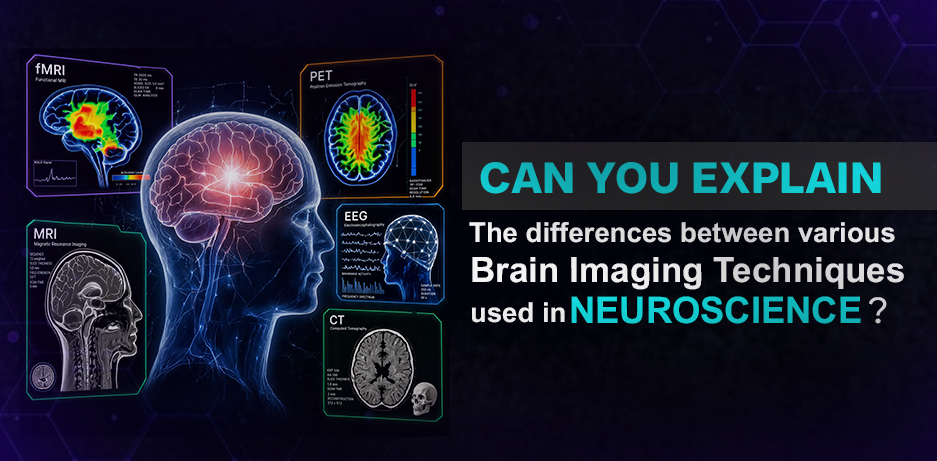
- CT (Computed Tomography): Uses X rays for fast cross-sectional images. It’s the go-to in emergencies for detecting bleeding, fractures, or large strokes.
- MRI (Magnetic Resonance Imaging): Uses magnetic fields and radio waves for high-resolution soft-tissue detail. Preferred for tumours, small lesions, and subtle structural changes, the same level of detail surgeons rely on when planning brain tumour surgery.
- DTI (Diffusion Tensor Imaging): A specialised MRI variant that tracks water movement along white matter tracts, mapping neural connections and detecting damage from injury or disease.
Functional Brain Imaging Techniques
Functional neuroimaging reveals how the brain works in real time, not just what it looks like.
- fMRI (Functional MRI) Measures changes in blood flow to identify active brain regions during specific tasks. Used clinically for presurgical mapping and in cognitive research.
- PET (Positron Emission Tomography) Uses a small radioactive tracer to measure metabolic activity and neurotransmitter levels. Useful for detecting early metabolic changes linked to Alzheimer’s disease.
- EEG (Electroencephalography) Records electrical activity from electrodes placed on the scalp. Excellent time resolution makes it the standard for diagnosing epilepsy and studying sleep disorders, which is why an EEG test in Mumbai is often the first investigation ordered when seizures are suspected.
- SPECT (Single Photon Emission Computed Tomography) Similar to PET but more accessible, often used to assess blood flow after stroke or seizure activity.
Advanced Neuroimaging: What’s Next
Advanced neuroimaging is moving toward higher resolution, greater portability, and smarter interpretation. Ultra high field MRI (7 Tesla and above) reveals detail invisible to standard scanners, while machine learning models are being trained to spot patterns in neuroimaging data that even experienced radiologists might miss. Portable, wearable EEG and NIRS devices are expanding access to brain monitoring outside hospitals, from sports concussion checks to at home sleep studies. Hybrid scanners like PET MRI now combine anatomical and metabolic data in a single session, blurring the line between structural and functional neuroimaging.
How to Choose the Right Brain Imaging Technique
The right neuroimaging technique depends on what your doctor is trying to find:
- Emergency or suspected bleeding: CT scan, for speed
- Detailed structural concerns (tumours, lesions, MS): MRI
- Seizure or epilepsy evaluation: EEG
- Suspected dementia or metabolic disorders: PET scan
- Pre-surgical brain mapping: fMRI, sometimes paired with DTI
Symptoms and history guide which technique is best, which is why a consultation typically comes before the scan is ordered rather than after.
Limitations and Considerations in Neuroimaging
No single neuroimaging technique tells the whole story. Structural scans can miss functional problems, and functional scans often lack the anatomical detail needed for surgical planning. Cost and accessibility remain real barriers, since PET and MEG scanners aren’t available everywhere. Radiation exposure is a consideration with CT and PET, so these are used judiciously in children and pregnant patients. Movement can distort MRI and fMRI results, and interpreting functional data takes skilled radiologists to avoid false positives.
Neuroimaging works best alongside patient history and physical examination, not as a standalone answer, which is why a consultation with a neuro physician is usually the first step in deciding which scan is actually needed.
Conclusion
Brain imaging techniques have moved neuroscience from inference to direct observation. Whether it’s a CT scan catching a bleed in the emergency room or an fMRI mapping activity before surgery, each neuroimaging technique has a specific role and limitation. Understanding these differences helps patients ask better questions and helps decision makers choose the right diagnostic path forward. As advanced neuroimaging continues to evolve, expect faster, more precise, and more accessible ways to look inside the brain.
FAQs
1. How has neuroimaging changed the field of neuroscience over the past 20 years?
It’s shifted neuroscience from theory driven research to evidence based observation, enabling real-time study of brain activity and earlier disease detection.
2. Can brain imaging predict neurological conditions before symptoms appear?
Sometimes. PET and advanced MRI can detect early metabolic or structural changes linked to conditions like Alzheimer’s, occasionally years before symptoms appear.
3. Is brain imaging safe for children and pregnant women?
MRI and EEG are generally safe since they don’t use radiation. CT and PET involve radiation exposure and are used only when clearly necessary.
4. How long does a typical neuroimaging session take from start to finish?
Most sessions run 15 minutes to an hour. A CT scan can take under 15 minutes, while a detailed MRI or fMRI session may take 45 minutes or more.
5. What training or qualifications does a technician need to operate brain imaging equipment?
Technicians typically need specialised radiologic or MRI certification and supervised clinical training, with scans interpreted by qualified radiologists or neurologists.